Fluorine »
PDB 2bcj-2dux »
2bub »
Fluorine in PDB 2bub: Crystal Structure of Human Dipeptidyl Peptidase IV (CD26) in Complex with A Reversed Amide Inhibitor
Enzymatic activity of Crystal Structure of Human Dipeptidyl Peptidase IV (CD26) in Complex with A Reversed Amide Inhibitor
All present enzymatic activity of Crystal Structure of Human Dipeptidyl Peptidase IV (CD26) in Complex with A Reversed Amide Inhibitor:
3.4.14.5;
3.4.14.5;
Protein crystallography data
The structure of Crystal Structure of Human Dipeptidyl Peptidase IV (CD26) in Complex with A Reversed Amide Inhibitor, PDB code: 2bub
was solved by
S.Nordhoff,
S.Cerezo-Galvez,
A.Feurer,
O.Hill,
V.G.Matassa,
G.Metz,
C.Rummey,
M.Thiemann,
P.J.Edwards,
with X-Ray Crystallography technique. A brief refinement statistics is given in the table below:
| Resolution Low / High (Å) | 20.00 / 2.66 |
| Space group | P 21 21 21 |
| Cell size a, b, c (Å), α, β, γ (°) | 65.486, 66.773, 425.407, 90.00, 90.00, 90.00 |
| R / Rfree (%) | 25.8 / 32.9 |
Fluorine Binding Sites:
The binding sites of Fluorine atom in the Crystal Structure of Human Dipeptidyl Peptidase IV (CD26) in Complex with A Reversed Amide Inhibitor
(pdb code 2bub). This binding sites where shown within
5.0 Angstroms radius around Fluorine atom.
In total 2 binding sites of Fluorine where determined in the Crystal Structure of Human Dipeptidyl Peptidase IV (CD26) in Complex with A Reversed Amide Inhibitor, PDB code: 2bub:
Jump to Fluorine binding site number: 1; 2;
In total 2 binding sites of Fluorine where determined in the Crystal Structure of Human Dipeptidyl Peptidase IV (CD26) in Complex with A Reversed Amide Inhibitor, PDB code: 2bub:
Jump to Fluorine binding site number: 1; 2;
Fluorine binding site 1 out of 2 in 2bub
Go back to
Fluorine binding site 1 out
of 2 in the Crystal Structure of Human Dipeptidyl Peptidase IV (CD26) in Complex with A Reversed Amide Inhibitor
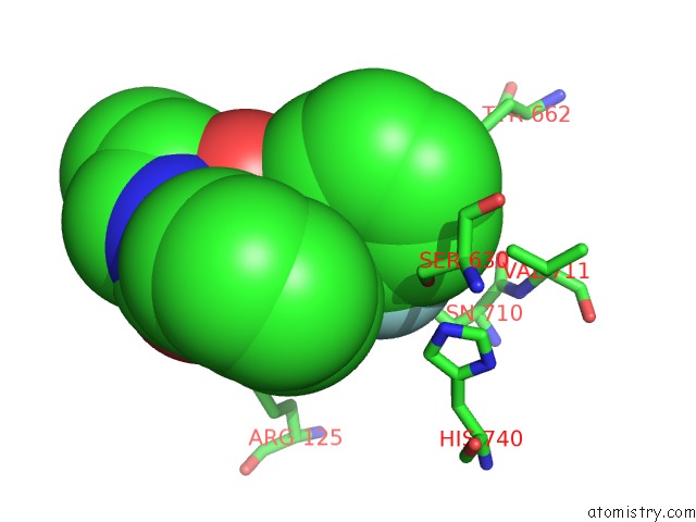
Mono view
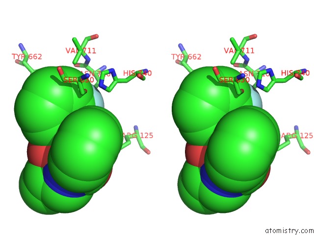
Stereo pair view
Mono view
Stereo pair view
A full contact list of Fluorine with other atoms in the F binding
site number 1 of Crystal Structure of Human Dipeptidyl Peptidase IV (CD26) in Complex with A Reversed Amide Inhibitor within 5.0Å range:
|
Fluorine binding site 2 out of 2 in 2bub
Go back to
Fluorine binding site 2 out
of 2 in the Crystal Structure of Human Dipeptidyl Peptidase IV (CD26) in Complex with A Reversed Amide Inhibitor
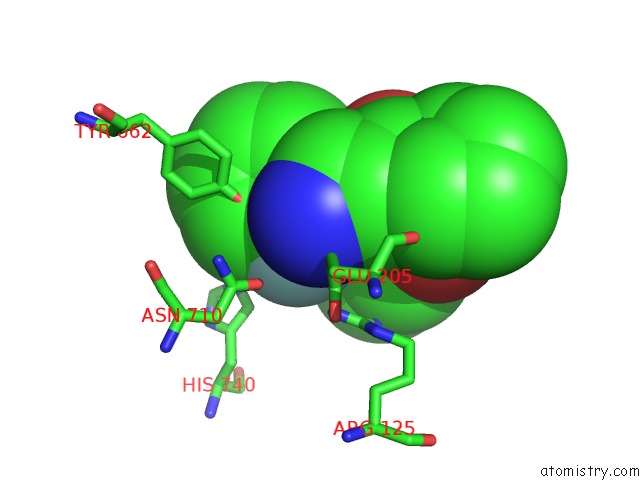
Mono view
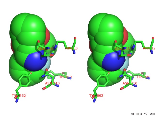
Stereo pair view
Mono view
Stereo pair view
A full contact list of Fluorine with other atoms in the F binding
site number 2 of Crystal Structure of Human Dipeptidyl Peptidase IV (CD26) in Complex with A Reversed Amide Inhibitor within 5.0Å range:
|
Reference:
S.Nordhoff,
S.Cerezo-Galvez,
A.Feurer,
O.Hill,
V.G.Matassa,
G.Metz,
C.Rummey,
M.Thiemann,
P.J.Edwards.
The Reversed Binding of Beta-Phenethylamine Inhibitors of Dpp-IV: X-Ray Structures and Properties of Novel Fragment and Elaborated Inhibitors. Bioorg. Med. Chem. Lett. V. 16 1744 2006.
ISSN: ISSN 0960-894X
PubMed: 16376544
DOI: 10.1016/J.BMCL.2005.11.103
Page generated: Wed Jul 31 13:53:19 2024
ISSN: ISSN 0960-894X
PubMed: 16376544
DOI: 10.1016/J.BMCL.2005.11.103
Last articles
Ca in 5NERCa in 5NEM
Ca in 5NE5
Ca in 5NBP
Ca in 5NBN
Ca in 5NBM
Ca in 5NBL
Ca in 5N7G
Ca in 5N7F
Ca in 5N7D