Fluorine »
PDB 2bcj-2dux »
2cmc »
Fluorine in PDB 2cmc: Structural Basis For Inhibition of Protein Tyrosine Phosphatase 1B By Isothiazolidinone Heterocyclic Phosphonate Mimetics
Enzymatic activity of Structural Basis For Inhibition of Protein Tyrosine Phosphatase 1B By Isothiazolidinone Heterocyclic Phosphonate Mimetics
All present enzymatic activity of Structural Basis For Inhibition of Protein Tyrosine Phosphatase 1B By Isothiazolidinone Heterocyclic Phosphonate Mimetics:
3.1.3.48;
3.1.3.48;
Protein crystallography data
The structure of Structural Basis For Inhibition of Protein Tyrosine Phosphatase 1B By Isothiazolidinone Heterocyclic Phosphonate Mimetics, PDB code: 2cmc
was solved by
P.J.Ala,
L.Gonneville,
M.C.Hillman,
M.Becker-Pasha,
M.Wei,
B.G.Reid,
R.Klabe,
E.W.Yue,
B.Wayland,
B.Douty,
A.P.Combs,
P.Polam,
Z.Wasserman,
M.Bower,
T.C.Burn,
G.F.Hollis,
R.Wynn,
with X-Ray Crystallography technique. A brief refinement statistics is given in the table below:
| Resolution Low / High (Å) | 7.00 / 2.20 |
| Space group | P 21 21 21 |
| Cell size a, b, c (Å), α, β, γ (°) | 62.200, 70.900, 83.310, 90.00, 90.00, 90.00 |
| R / Rfree (%) | n/a / n/a |
Fluorine Binding Sites:
The binding sites of Fluorine atom in the Structural Basis For Inhibition of Protein Tyrosine Phosphatase 1B By Isothiazolidinone Heterocyclic Phosphonate Mimetics
(pdb code 2cmc). This binding sites where shown within
5.0 Angstroms radius around Fluorine atom.
In total 2 binding sites of Fluorine where determined in the Structural Basis For Inhibition of Protein Tyrosine Phosphatase 1B By Isothiazolidinone Heterocyclic Phosphonate Mimetics, PDB code: 2cmc:
Jump to Fluorine binding site number: 1; 2;
In total 2 binding sites of Fluorine where determined in the Structural Basis For Inhibition of Protein Tyrosine Phosphatase 1B By Isothiazolidinone Heterocyclic Phosphonate Mimetics, PDB code: 2cmc:
Jump to Fluorine binding site number: 1; 2;
Fluorine binding site 1 out of 2 in 2cmc
Go back to
Fluorine binding site 1 out
of 2 in the Structural Basis For Inhibition of Protein Tyrosine Phosphatase 1B By Isothiazolidinone Heterocyclic Phosphonate Mimetics
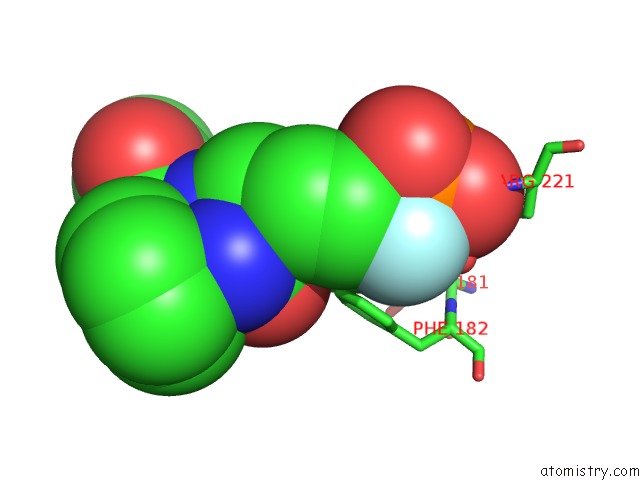
Mono view
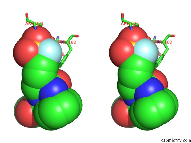
Stereo pair view
Mono view
Stereo pair view
A full contact list of Fluorine with other atoms in the F binding
site number 1 of Structural Basis For Inhibition of Protein Tyrosine Phosphatase 1B By Isothiazolidinone Heterocyclic Phosphonate Mimetics within 5.0Å range:
|
Fluorine binding site 2 out of 2 in 2cmc
Go back to
Fluorine binding site 2 out
of 2 in the Structural Basis For Inhibition of Protein Tyrosine Phosphatase 1B By Isothiazolidinone Heterocyclic Phosphonate Mimetics
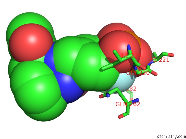
Mono view
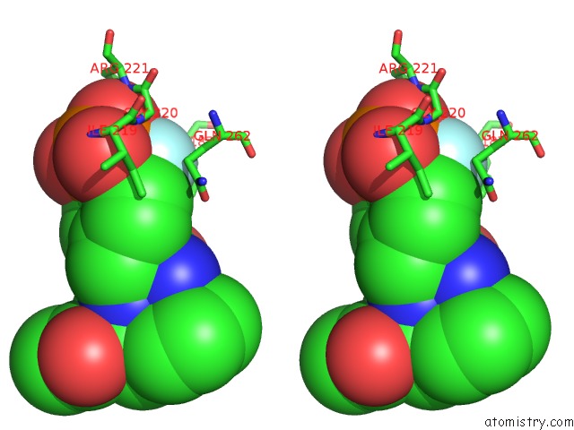
Stereo pair view
Mono view
Stereo pair view
A full contact list of Fluorine with other atoms in the F binding
site number 2 of Structural Basis For Inhibition of Protein Tyrosine Phosphatase 1B By Isothiazolidinone Heterocyclic Phosphonate Mimetics within 5.0Å range:
|
Reference:
P.J.Ala,
L.Gonneville,
M.C.Hillman,
M.Becker-Pasha,
M.Wei,
B.G.Reid,
R.Klabe,
E.W.Yue,
B.Wayland,
B.Douty,
P.Polam,
Z.Wasserman,
M.Bower,
A.P.Combs,
T.C.Burn,
G.F.Hollis,
R.Wynn.
Structural Basis For Inhibition of Protein-Tyrosine Phosphatase 1B By Isothiazolidinone Heterocyclic Phosphonate Mimetics. J.Biol.Chem. V. 281 32784 2006.
ISSN: ISSN 0021-9258
PubMed: 16916797
DOI: 10.1074/JBC.M606873200
Page generated: Wed Jul 31 13:59:29 2024
ISSN: ISSN 0021-9258
PubMed: 16916797
DOI: 10.1074/JBC.M606873200
Last articles
Cl in 5U8JCl in 5U8O
Cl in 5U81
Cl in 5U8G
Cl in 5U72
Cl in 5U7K
Cl in 5U86
Cl in 5U6X
Cl in 5U6W
Cl in 5U6B