Fluorine »
PDB 2bcj-2dux »
2dqz »
Fluorine in PDB 2dqz: Crystal Structure of Human Carboxylesterase in Complex with Homatropine, Coenzyme A, and Palmitate
Enzymatic activity of Crystal Structure of Human Carboxylesterase in Complex with Homatropine, Coenzyme A, and Palmitate
All present enzymatic activity of Crystal Structure of Human Carboxylesterase in Complex with Homatropine, Coenzyme A, and Palmitate:
3.1.1.1;
3.1.1.1;
Protein crystallography data
The structure of Crystal Structure of Human Carboxylesterase in Complex with Homatropine, Coenzyme A, and Palmitate, PDB code: 2dqz
was solved by
S.Bencharit,
M.R.Redinbo,
with X-Ray Crystallography technique. A brief refinement statistics is given in the table below:
| Resolution Low / High (Å) | 22.60 / 2.80 |
| Space group | P 21 21 21 |
| Cell size a, b, c (Å), α, β, γ (°) | 55.564, 181.017, 202.560, 90.00, 90.00, 90.00 |
| R / Rfree (%) | 19.3 / 24.4 |
Fluorine Binding Sites:
The binding sites of Fluorine atom in the Crystal Structure of Human Carboxylesterase in Complex with Homatropine, Coenzyme A, and Palmitate
(pdb code 2dqz). This binding sites where shown within
5.0 Angstroms radius around Fluorine atom.
In total 3 binding sites of Fluorine where determined in the Crystal Structure of Human Carboxylesterase in Complex with Homatropine, Coenzyme A, and Palmitate, PDB code: 2dqz:
Jump to Fluorine binding site number: 1; 2; 3;
In total 3 binding sites of Fluorine where determined in the Crystal Structure of Human Carboxylesterase in Complex with Homatropine, Coenzyme A, and Palmitate, PDB code: 2dqz:
Jump to Fluorine binding site number: 1; 2; 3;
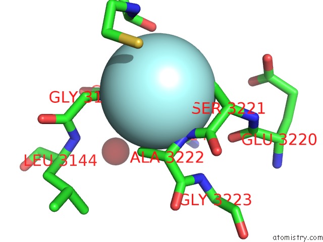
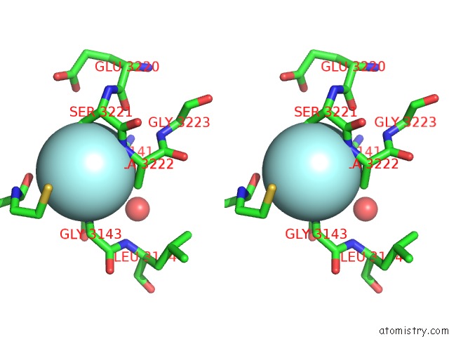
Fluorine binding site 1 out of 3 in 2dqz
Go back to
Fluorine binding site 1 out
of 3 in the Crystal Structure of Human Carboxylesterase in Complex with Homatropine, Coenzyme A, and Palmitate
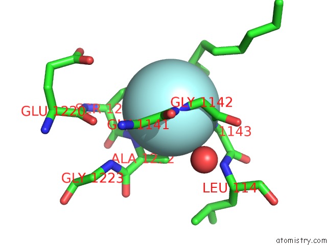
Mono view
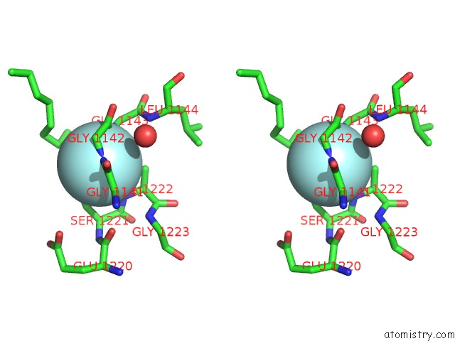
Stereo pair view
Mono view
Stereo pair view
A full contact list of Fluorine with other atoms in the F binding
site number 1 of Crystal Structure of Human Carboxylesterase in Complex with Homatropine, Coenzyme A, and Palmitate within 5.0Å range:
|
Fluorine binding site 2 out of 3 in 2dqz
Go back to
Fluorine binding site 2 out
of 3 in the Crystal Structure of Human Carboxylesterase in Complex with Homatropine, Coenzyme A, and Palmitate
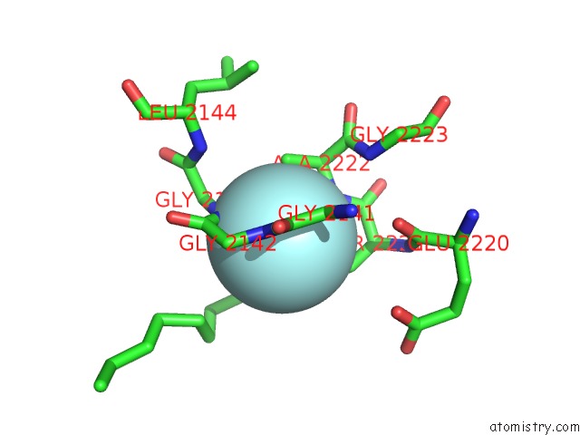
Mono view
Stereo pair view
Mono view
Stereo pair view
A full contact list of Fluorine with other atoms in the F binding
site number 2 of Crystal Structure of Human Carboxylesterase in Complex with Homatropine, Coenzyme A, and Palmitate within 5.0Å range:
|
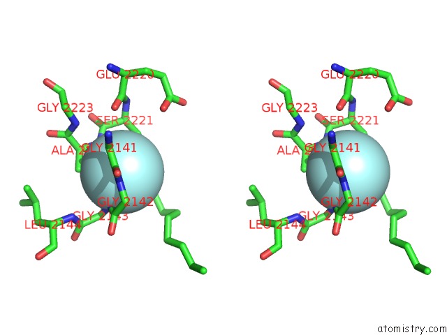
Fluorine binding site 3 out of 3 in 2dqz
Go back to
Fluorine binding site 3 out
of 3 in the Crystal Structure of Human Carboxylesterase in Complex with Homatropine, Coenzyme A, and Palmitate
Mono view
Stereo pair view
Mono view
Stereo pair view
A full contact list of Fluorine with other atoms in the F binding
site number 3 of Crystal Structure of Human Carboxylesterase in Complex with Homatropine, Coenzyme A, and Palmitate within 5.0Å range:
|
Reference:
S.Bencharit,
C.C.Edwards,
C.L.Morton,
E.L.Howard-Williams,
P.Kuhn,
P.M.Potter,
M.R.Redinbo.
Multisite Promiscuity in the Processing of Endogenous Substrates By Human Carboxylesterase 1 J.Mol.Biol. V. 363 201 2006.
ISSN: ISSN 0022-2836
PubMed: 16962139
DOI: 10.1016/J.JMB.2006.08.025
Page generated: Wed Jul 31 14:02:46 2024
ISSN: ISSN 0022-2836
PubMed: 16962139
DOI: 10.1016/J.JMB.2006.08.025
Last articles
Zn in 9J0NZn in 9J0O
Zn in 9J0P
Zn in 9FJX
Zn in 9EKB
Zn in 9C0F
Zn in 9CAH
Zn in 9CH0
Zn in 9CH3
Zn in 9CH1