Fluorine »
PDB 2bcj-2dux »
2drj »
Fluorine in PDB 2drj: Xray Structure of Alpha-2,3/8-Sialyltransferase Cstii F91Y Mutant
Protein crystallography data
The structure of Xray Structure of Alpha-2,3/8-Sialyltransferase Cstii F91Y Mutant, PDB code: 2drj
was solved by
C.P.C.Chiu,
N.C.J.Strynadka,
with X-Ray Crystallography technique. A brief refinement statistics is given in the table below:
| Resolution Low / High (Å) | 23.81 / 2.25 |
| Space group | I 4 |
| Cell size a, b, c (Å), α, β, γ (°) | 116.653, 116.653, 44.086, 90.00, 90.00, 90.00 |
| R / Rfree (%) | 19.4 / 26.1 |
Fluorine Binding Sites:
The binding sites of Fluorine atom in the Xray Structure of Alpha-2,3/8-Sialyltransferase Cstii F91Y Mutant
(pdb code 2drj). This binding sites where shown within
5.0 Angstroms radius around Fluorine atom.
In total only one binding site of Fluorine was determined in the Xray Structure of Alpha-2,3/8-Sialyltransferase Cstii F91Y Mutant, PDB code: 2drj:
In total only one binding site of Fluorine was determined in the Xray Structure of Alpha-2,3/8-Sialyltransferase Cstii F91Y Mutant, PDB code: 2drj:
Fluorine binding site 1 out of 1 in 2drj
Go back to
Fluorine binding site 1 out
of 1 in the Xray Structure of Alpha-2,3/8-Sialyltransferase Cstii F91Y Mutant
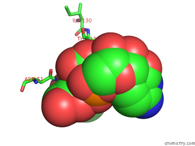
Mono view
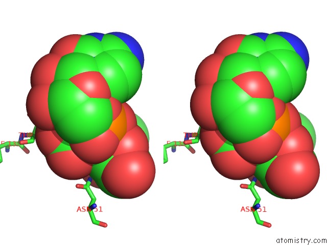
Stereo pair view
Mono view
Stereo pair view
A full contact list of Fluorine with other atoms in the F binding
site number 1 of Xray Structure of Alpha-2,3/8-Sialyltransferase Cstii F91Y Mutant within 5.0Å range:
|
Reference:
A.Aharoni,
K.Thieme,
C.P.C.Chiu,
S.Buchini,
L.L.Lairson,
H.Chen,
N.C.J.Strynadka,
W.W.Wakarchuk,
S.G.Withers.
High-Throughput Screening Methodology For the Directed Evolution of Glycosyltransferases Nat.Methods V. 3 609 2006.
ISSN: ISSN 1548-7091
PubMed: 16862130
DOI: 10.1038/NMETH899
Page generated: Mon Jul 14 12:50:14 2025
ISSN: ISSN 1548-7091
PubMed: 16862130
DOI: 10.1038/NMETH899
Last articles
F in 7HB6F in 7HAP
F in 7H7I
F in 7H73
F in 7H75
F in 7H6V
F in 7H6W
F in 7H5S
F in 7H5R
F in 7H5T