Fluorine »
PDB 2duz-2fq6 »
2eud »
Fluorine in PDB 2eud: Structures of Yeast Ribonucleotide Reductase I Complexed with Ligands and Subunit Peptides
Enzymatic activity of Structures of Yeast Ribonucleotide Reductase I Complexed with Ligands and Subunit Peptides
All present enzymatic activity of Structures of Yeast Ribonucleotide Reductase I Complexed with Ligands and Subunit Peptides:
1.17.4.1;
1.17.4.1;
Protein crystallography data
The structure of Structures of Yeast Ribonucleotide Reductase I Complexed with Ligands and Subunit Peptides, PDB code: 2eud
was solved by
C.Dealwis,
H.Xu,
C.Faber,
T.Uchiki,
J.W.Fairman,
J.Racca,
with X-Ray Crystallography technique. A brief refinement statistics is given in the table below:
| Resolution Low / High (Å) | 50.00 / 2.30 |
| Space group | P 21 21 2 |
| Cell size a, b, c (Å), α, β, γ (°) | 107.578, 117.216, 63.990, 90.00, 90.00, 90.00 |
| R / Rfree (%) | 20.4 / 24 |
Other elements in 2eud:
The structure of Structures of Yeast Ribonucleotide Reductase I Complexed with Ligands and Subunit Peptides also contains other interesting chemical elements:
| Magnesium | (Mg) | 1 atom |
Fluorine Binding Sites:
The binding sites of Fluorine atom in the Structures of Yeast Ribonucleotide Reductase I Complexed with Ligands and Subunit Peptides
(pdb code 2eud). This binding sites where shown within
5.0 Angstroms radius around Fluorine atom.
In total 2 binding sites of Fluorine where determined in the Structures of Yeast Ribonucleotide Reductase I Complexed with Ligands and Subunit Peptides, PDB code: 2eud:
Jump to Fluorine binding site number: 1; 2;
In total 2 binding sites of Fluorine where determined in the Structures of Yeast Ribonucleotide Reductase I Complexed with Ligands and Subunit Peptides, PDB code: 2eud:
Jump to Fluorine binding site number: 1; 2;
Fluorine binding site 1 out of 2 in 2eud
Go back to
Fluorine binding site 1 out
of 2 in the Structures of Yeast Ribonucleotide Reductase I Complexed with Ligands and Subunit Peptides
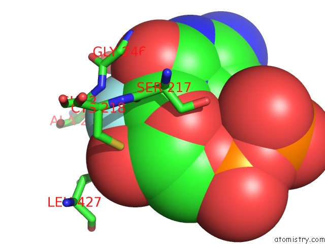
Mono view
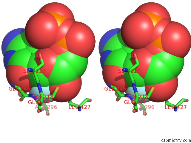
Stereo pair view
Mono view
Stereo pair view
A full contact list of Fluorine with other atoms in the F binding
site number 1 of Structures of Yeast Ribonucleotide Reductase I Complexed with Ligands and Subunit Peptides within 5.0Å range:
|
Fluorine binding site 2 out of 2 in 2eud
Go back to
Fluorine binding site 2 out
of 2 in the Structures of Yeast Ribonucleotide Reductase I Complexed with Ligands and Subunit Peptides
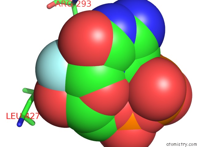
Mono view
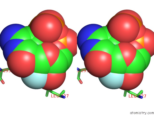
Stereo pair view
Mono view
Stereo pair view
A full contact list of Fluorine with other atoms in the F binding
site number 2 of Structures of Yeast Ribonucleotide Reductase I Complexed with Ligands and Subunit Peptides within 5.0Å range:
|
Reference:
H.Xu,
C.Faber,
T.Uchiki,
J.Racca,
C.Dealwis.
Structures of Eukaryotic Ribonucleotide Reductase I Define Gemcitabine Diphosphate Binding and Subunit Assembly. Proc.Natl.Acad.Sci.Usa V. 103 4028 2006.
ISSN: ISSN 0027-8424
PubMed: 16537480
DOI: 10.1073/PNAS.0600440103
Page generated: Wed Jul 31 14:07:30 2024
ISSN: ISSN 0027-8424
PubMed: 16537480
DOI: 10.1073/PNAS.0600440103
Last articles
Zn in 9MJ5Zn in 9HNW
Zn in 9G0L
Zn in 9FNE
Zn in 9DZN
Zn in 9E0I
Zn in 9D32
Zn in 9DAK
Zn in 8ZXC
Zn in 8ZUF