Fluorine »
PDB 2duz-2fq6 »
2fjn »
Fluorine in PDB 2fjn: The Structure of Phosphotyrosine Phosphatase 1B in Complex with Compound 2
Enzymatic activity of The Structure of Phosphotyrosine Phosphatase 1B in Complex with Compound 2
All present enzymatic activity of The Structure of Phosphotyrosine Phosphatase 1B in Complex with Compound 2:
3.1.3.48;
3.1.3.48;
Protein crystallography data
The structure of The Structure of Phosphotyrosine Phosphatase 1B in Complex with Compound 2, PDB code: 2fjn
was solved by
E.Asante-Appiah,
S.Patel,
C.Desponts,
J.M.Taylor,
C.Lau,
C.Dufresne,
M.Therien,
R.Friesen,
J.W.Becker,
Y.Leblanc,
B.P.Kennedy,
G.Scapin,
with X-Ray Crystallography technique. A brief refinement statistics is given in the table below:
| Resolution Low / High (Å) | 13.00 / 2.20 |
| Space group | P 21 21 21 |
| Cell size a, b, c (Å), α, β, γ (°) | 87.429, 85.617, 137.272, 90.00, 90.00, 90.00 |
| R / Rfree (%) | n/a / n/a |
Other elements in 2fjn:
The structure of The Structure of Phosphotyrosine Phosphatase 1B in Complex with Compound 2 also contains other interesting chemical elements:
| Chlorine | (Cl) | 2 atoms |
Fluorine Binding Sites:
The binding sites of Fluorine atom in the The Structure of Phosphotyrosine Phosphatase 1B in Complex with Compound 2
(pdb code 2fjn). This binding sites where shown within
5.0 Angstroms radius around Fluorine atom.
In total 4 binding sites of Fluorine where determined in the The Structure of Phosphotyrosine Phosphatase 1B in Complex with Compound 2, PDB code: 2fjn:
Jump to Fluorine binding site number: 1; 2; 3; 4;
In total 4 binding sites of Fluorine where determined in the The Structure of Phosphotyrosine Phosphatase 1B in Complex with Compound 2, PDB code: 2fjn:
Jump to Fluorine binding site number: 1; 2; 3; 4;
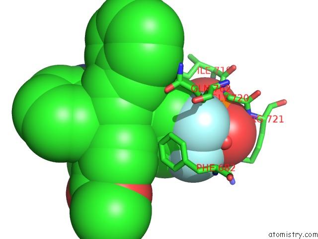
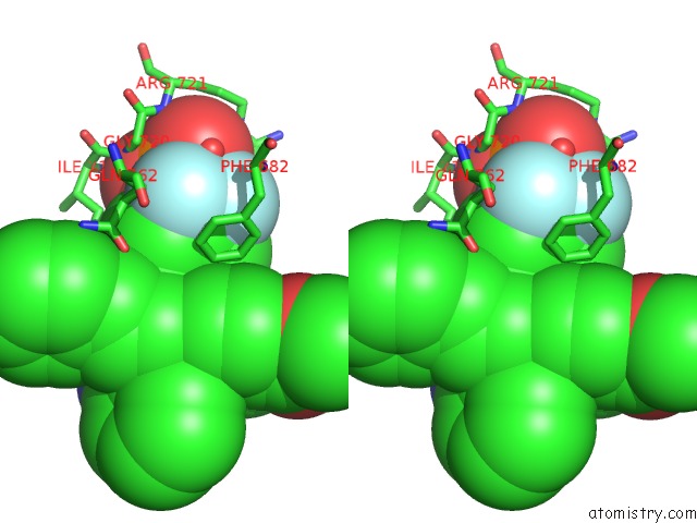
Fluorine binding site 1 out of 4 in 2fjn
Go back to
Fluorine binding site 1 out
of 4 in the The Structure of Phosphotyrosine Phosphatase 1B in Complex with Compound 2
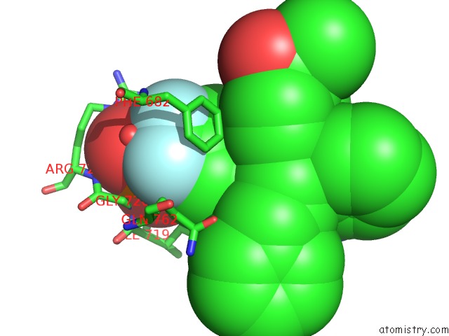
Mono view
Stereo pair view
Mono view
Stereo pair view
A full contact list of Fluorine with other atoms in the F binding
site number 1 of The Structure of Phosphotyrosine Phosphatase 1B in Complex with Compound 2 within 5.0Å range:
|
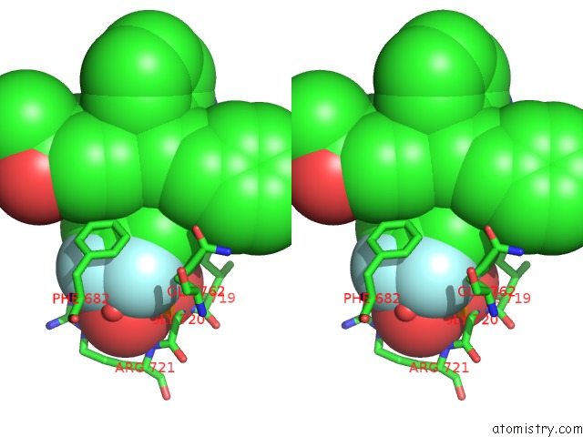
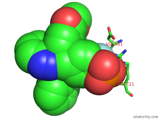
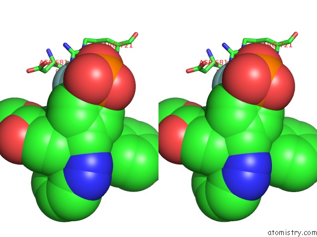
Fluorine binding site 2 out of 4 in 2fjn
Go back to
Fluorine binding site 2 out
of 4 in the The Structure of Phosphotyrosine Phosphatase 1B in Complex with Compound 2
Mono view
Stereo pair view
Mono view
Stereo pair view
A full contact list of Fluorine with other atoms in the F binding
site number 2 of The Structure of Phosphotyrosine Phosphatase 1B in Complex with Compound 2 within 5.0Å range:
|
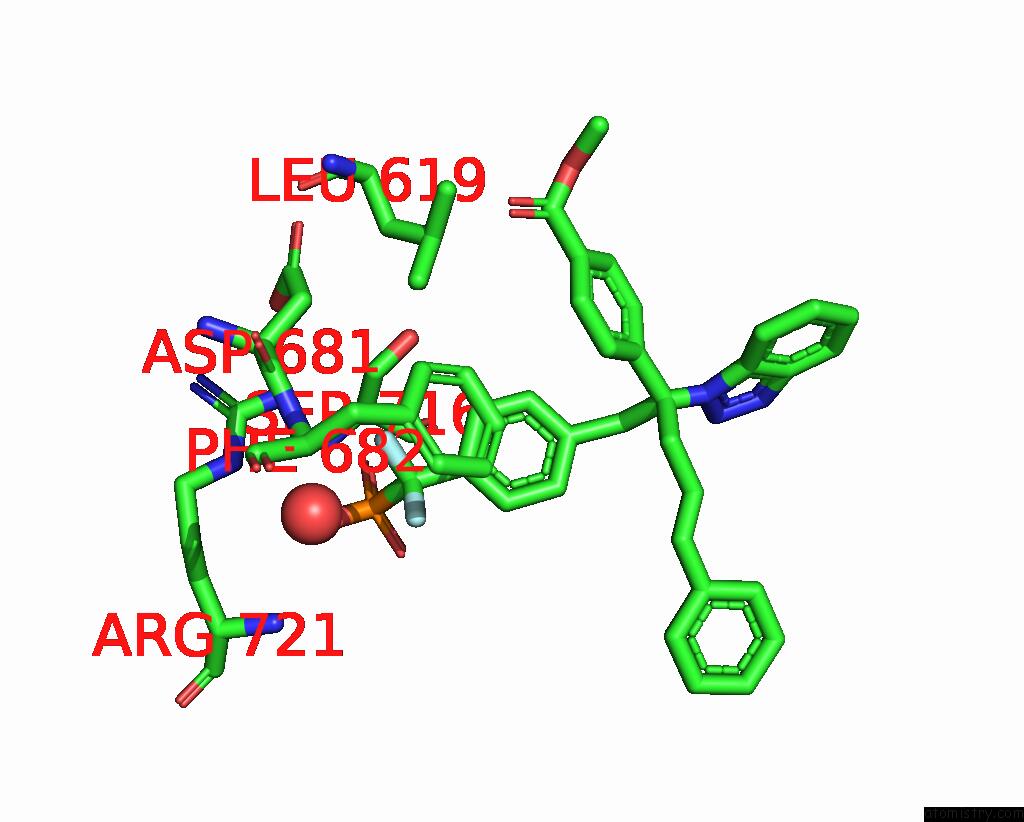
Fluorine binding site 3 out of 4 in 2fjn
Go back to
Fluorine binding site 3 out
of 4 in the The Structure of Phosphotyrosine Phosphatase 1B in Complex with Compound 2
Mono view
Stereo pair view
Mono view
Stereo pair view
A full contact list of Fluorine with other atoms in the F binding
site number 3 of The Structure of Phosphotyrosine Phosphatase 1B in Complex with Compound 2 within 5.0Å range:
|
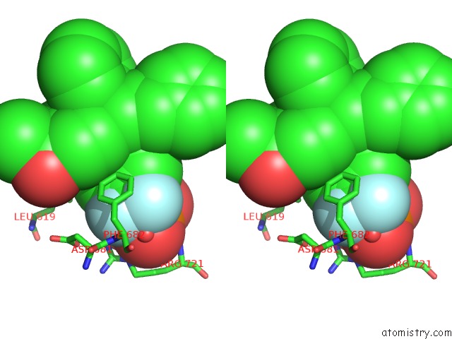
Fluorine binding site 4 out of 4 in 2fjn
Go back to
Fluorine binding site 4 out
of 4 in the The Structure of Phosphotyrosine Phosphatase 1B in Complex with Compound 2
Mono view
Stereo pair view
Mono view
Stereo pair view
A full contact list of Fluorine with other atoms in the F binding
site number 4 of The Structure of Phosphotyrosine Phosphatase 1B in Complex with Compound 2 within 5.0Å range:
|
Reference:
E.Asante-Appiah,
S.Patel,
C.Desponts,
J.M.Taylor,
C.Lau,
C.Dufresne,
M.Therien,
R.Friesen,
J.W.Becker,
Y.Leblanc,
B.P.Kennedy,
G.Scapin.
Conformation-Assisted Inhibition of Protein-Tyrosine Phosphatase-1B Elicits Inhibitor Selectivity Over T-Cell Protein-Tyrosine Phosphatase. J.Biol.Chem. V. 281 8010 2006.
ISSN: ISSN 0021-9258
PubMed: 16407290
DOI: 10.1074/JBC.M511827200
Page generated: Mon Jul 14 12:56:22 2025
ISSN: ISSN 0021-9258
PubMed: 16407290
DOI: 10.1074/JBC.M511827200
Last articles
Fe in 2YXOFe in 2YRS
Fe in 2YXC
Fe in 2YNM
Fe in 2YVJ
Fe in 2YP1
Fe in 2YU2
Fe in 2YU1
Fe in 2YQB
Fe in 2YOO