Fluorine »
PDB 2gtn-2ihj »
2i0h »
Fluorine in PDB 2i0h: The Structure of P38ALPHA in Complex with An Arylpyridazinone
Enzymatic activity of The Structure of P38ALPHA in Complex with An Arylpyridazinone
All present enzymatic activity of The Structure of P38ALPHA in Complex with An Arylpyridazinone:
2.7.11.24;
2.7.11.24;
Protein crystallography data
The structure of The Structure of P38ALPHA in Complex with An Arylpyridazinone, PDB code: 2i0h
was solved by
S.R.Natarajan,
S.T.Heller,
K.Nam,
S.B.Singh,
G.Scapin,
S.Patel,
J.E.Thompson,
C.E.Fitzgerald,
S.J.O'keefe,
with X-Ray Crystallography technique. A brief refinement statistics is given in the table below:
| Resolution Low / High (Å) | 30.00 / 2.00 |
| Space group | P 21 21 21 |
| Cell size a, b, c (Å), α, β, γ (°) | 44.722, 88.225, 121.456, 90.00, 90.00, 90.00 |
| R / Rfree (%) | 17.8 / 22.8 |
Other elements in 2i0h:
The structure of The Structure of P38ALPHA in Complex with An Arylpyridazinone also contains other interesting chemical elements:
| Chlorine | (Cl) | 4 atoms |
Fluorine Binding Sites:
The binding sites of Fluorine atom in the The Structure of P38ALPHA in Complex with An Arylpyridazinone
(pdb code 2i0h). This binding sites where shown within
5.0 Angstroms radius around Fluorine atom.
In total 2 binding sites of Fluorine where determined in the The Structure of P38ALPHA in Complex with An Arylpyridazinone, PDB code: 2i0h:
Jump to Fluorine binding site number: 1; 2;
In total 2 binding sites of Fluorine where determined in the The Structure of P38ALPHA in Complex with An Arylpyridazinone, PDB code: 2i0h:
Jump to Fluorine binding site number: 1; 2;
Fluorine binding site 1 out of 2 in 2i0h
Go back to
Fluorine binding site 1 out
of 2 in the The Structure of P38ALPHA in Complex with An Arylpyridazinone
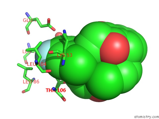
Mono view
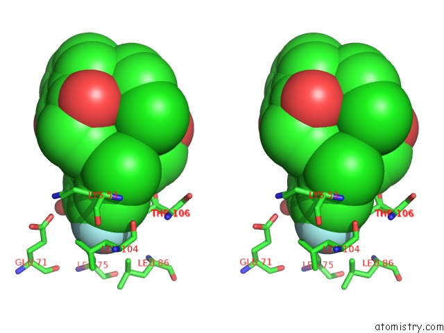
Stereo pair view
Mono view
Stereo pair view
A full contact list of Fluorine with other atoms in the F binding
site number 1 of The Structure of P38ALPHA in Complex with An Arylpyridazinone within 5.0Å range:
|
Fluorine binding site 2 out of 2 in 2i0h
Go back to
Fluorine binding site 2 out
of 2 in the The Structure of P38ALPHA in Complex with An Arylpyridazinone
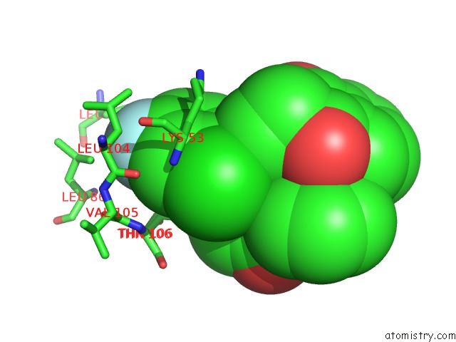
Mono view
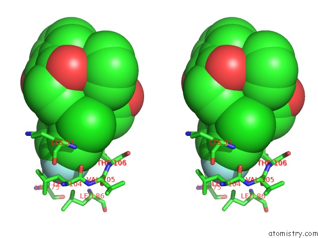
Stereo pair view
Mono view
Stereo pair view
A full contact list of Fluorine with other atoms in the F binding
site number 2 of The Structure of P38ALPHA in Complex with An Arylpyridazinone within 5.0Å range:
|
Reference:
S.R.Natarajan,
S.T.Heller,
K.Nam,
S.B.Singh,
G.Scapin,
S.Patel,
C.E.Fitzgerald,
J.E.Thompson,
S.J.O'keefe.
P38 Map Kinase Inhibitors Part 6: 2-Arylpyridazin-3-Ones As Templates For Inhibitor Design. Bioorg.Med.Chem.Lett. V. 16 5809 2006.
ISSN: ISSN 0960-894X
PubMed: 16945533
DOI: 10.1016/J.BMCL.2006.08.074
Page generated: Wed Jul 31 14:48:33 2024
ISSN: ISSN 0960-894X
PubMed: 16945533
DOI: 10.1016/J.BMCL.2006.08.074
Last articles
Ca in 5XQ3Ca in 5XPX
Ca in 5XP3
Ca in 5XPS
Ca in 5XNQ
Ca in 5XNR
Ca in 5XNM
Ca in 5XNL
Ca in 5XND
Ca in 5XLZ