Fluorine »
PDB 2gtn-2ihj »
2i82 »
Fluorine in PDB 2i82: Crystal Structure of Pseudouridine Synthase Rlua: Indirect Sequence Readout Through Protein-Induced Rna Structure
Enzymatic activity of Crystal Structure of Pseudouridine Synthase Rlua: Indirect Sequence Readout Through Protein-Induced Rna Structure
All present enzymatic activity of Crystal Structure of Pseudouridine Synthase Rlua: Indirect Sequence Readout Through Protein-Induced Rna Structure:
4.2.1.70;
4.2.1.70;
Protein crystallography data
The structure of Crystal Structure of Pseudouridine Synthase Rlua: Indirect Sequence Readout Through Protein-Induced Rna Structure, PDB code: 2i82
was solved by
C.Hoang,
with X-Ray Crystallography technique. A brief refinement statistics is given in the table below:
| Resolution Low / High (Å) | 19.83 / 2.05 |
| Space group | C 1 2 1 |
| Cell size a, b, c (Å), α, β, γ (°) | 320.737, 51.691, 81.233, 90.00, 90.81, 90.00 |
| R / Rfree (%) | 23.4 / 26.3 |
Fluorine Binding Sites:
The binding sites of Fluorine atom in the Crystal Structure of Pseudouridine Synthase Rlua: Indirect Sequence Readout Through Protein-Induced Rna Structure
(pdb code 2i82). This binding sites where shown within
5.0 Angstroms radius around Fluorine atom.
In total 2 binding sites of Fluorine where determined in the Crystal Structure of Pseudouridine Synthase Rlua: Indirect Sequence Readout Through Protein-Induced Rna Structure, PDB code: 2i82:
Jump to Fluorine binding site number: 1; 2;
In total 2 binding sites of Fluorine where determined in the Crystal Structure of Pseudouridine Synthase Rlua: Indirect Sequence Readout Through Protein-Induced Rna Structure, PDB code: 2i82:
Jump to Fluorine binding site number: 1; 2;
Fluorine binding site 1 out of 2 in 2i82
Go back to
Fluorine binding site 1 out
of 2 in the Crystal Structure of Pseudouridine Synthase Rlua: Indirect Sequence Readout Through Protein-Induced Rna Structure
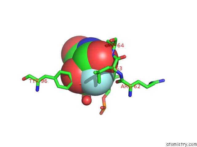
Mono view
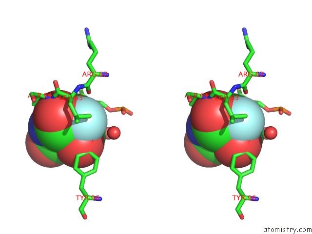
Stereo pair view
Mono view
Stereo pair view
A full contact list of Fluorine with other atoms in the F binding
site number 1 of Crystal Structure of Pseudouridine Synthase Rlua: Indirect Sequence Readout Through Protein-Induced Rna Structure within 5.0Å range:
|
Fluorine binding site 2 out of 2 in 2i82
Go back to
Fluorine binding site 2 out
of 2 in the Crystal Structure of Pseudouridine Synthase Rlua: Indirect Sequence Readout Through Protein-Induced Rna Structure
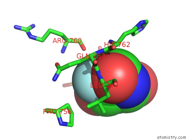
Mono view
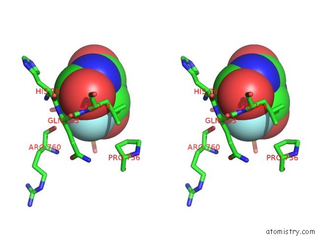
Stereo pair view
Mono view
Stereo pair view
A full contact list of Fluorine with other atoms in the F binding
site number 2 of Crystal Structure of Pseudouridine Synthase Rlua: Indirect Sequence Readout Through Protein-Induced Rna Structure within 5.0Å range:
|
Reference:
C.Hoang,
J.Chen,
C.A.Vizthum,
J.M.Kandel,
C.S.Hamilton,
E.G.Mueller,
A.R.Ferre-D'amare.
Crystal Structure of Pseudouridine Synthase Rlua: Indirect Sequence Readout Through Protein-Induced Rna Structure Mol.Cell V. 24 535 2006.
ISSN: ISSN 1097-2765
PubMed: 17188032
DOI: 10.1016/J.MOLCEL.2006.09.017
Page generated: Wed Jul 31 14:51:17 2024
ISSN: ISSN 1097-2765
PubMed: 17188032
DOI: 10.1016/J.MOLCEL.2006.09.017
Last articles
Zn in 9JYWZn in 9IR4
Zn in 9IR3
Zn in 9GMX
Zn in 9GMW
Zn in 9JEJ
Zn in 9ERF
Zn in 9ERE
Zn in 9EGV
Zn in 9EGW