Fluorine »
PDB 2ihk-2jxr »
2iko »
Fluorine in PDB 2iko: Crystal Structure of Human Renin Complexed with Inhibitor
Enzymatic activity of Crystal Structure of Human Renin Complexed with Inhibitor
All present enzymatic activity of Crystal Structure of Human Renin Complexed with Inhibitor:
3.4.23.15;
3.4.23.15;
Protein crystallography data
The structure of Crystal Structure of Human Renin Complexed with Inhibitor, PDB code: 2iko
was solved by
I.Mochalkin,
with X-Ray Crystallography technique. A brief refinement statistics is given in the table below:
| Resolution Low / High (Å) | 20.00 / 1.90 |
| Space group | P 21 3 |
| Cell size a, b, c (Å), α, β, γ (°) | 140.885, 140.885, 140.885, 90.00, 90.00, 90.00 |
| R / Rfree (%) | 19.4 / 22.9 |
Fluorine Binding Sites:
The binding sites of Fluorine atom in the Crystal Structure of Human Renin Complexed with Inhibitor
(pdb code 2iko). This binding sites where shown within
5.0 Angstroms radius around Fluorine atom.
In total 2 binding sites of Fluorine where determined in the Crystal Structure of Human Renin Complexed with Inhibitor, PDB code: 2iko:
Jump to Fluorine binding site number: 1; 2;
In total 2 binding sites of Fluorine where determined in the Crystal Structure of Human Renin Complexed with Inhibitor, PDB code: 2iko:
Jump to Fluorine binding site number: 1; 2;
Fluorine binding site 1 out of 2 in 2iko
Go back to
Fluorine binding site 1 out
of 2 in the Crystal Structure of Human Renin Complexed with Inhibitor

Mono view
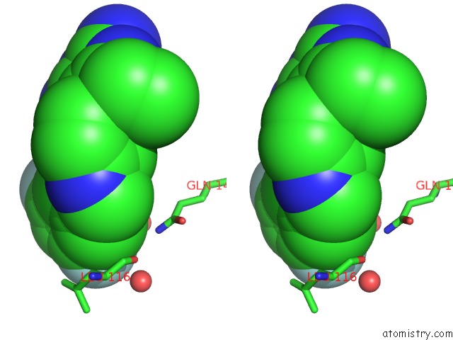
Stereo pair view
Mono view
Stereo pair view
A full contact list of Fluorine with other atoms in the F binding
site number 1 of Crystal Structure of Human Renin Complexed with Inhibitor within 5.0Å range:
|
Fluorine binding site 2 out of 2 in 2iko
Go back to
Fluorine binding site 2 out
of 2 in the Crystal Structure of Human Renin Complexed with Inhibitor
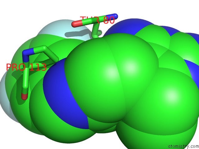
Mono view

Stereo pair view
Mono view
Stereo pair view
A full contact list of Fluorine with other atoms in the F binding
site number 2 of Crystal Structure of Human Renin Complexed with Inhibitor within 5.0Å range:
|
Reference:
R.W.Sarver,
J.Peevers,
W.L.Cody,
F.L.Ciske,
J.Dyer,
S.D.Emerson,
J.C.Hagadorn,
D.D.Holsworth,
M.Jalaie,
M.Kaufman,
M.Mastronardi,
P.Mcconnell,
N.A.Powell,
J.Quin,
C.A.Van Huis,
E.Zhang,
I.Mochalkin.
Binding Thermodynamics of Substituted Diaminopyrimidine Renin Inhibitors. Anal.Biochem. V. 360 30 2007.
ISSN: ISSN 0003-2697
PubMed: 17113558
DOI: 10.1016/J.AB.2006.10.017
Page generated: Wed Jul 31 14:54:33 2024
ISSN: ISSN 0003-2697
PubMed: 17113558
DOI: 10.1016/J.AB.2006.10.017
Last articles
Zn in 9J0NZn in 9J0O
Zn in 9J0P
Zn in 9FJX
Zn in 9EKB
Zn in 9C0F
Zn in 9CAH
Zn in 9CH0
Zn in 9CH3
Zn in 9CH1