Fluorine »
PDB 2ihk-2jxr »
2itz »
Fluorine in PDB 2itz: Crystal Structure of Egfr Kinase Domain L858R Mutation in Complex with Iressa
Enzymatic activity of Crystal Structure of Egfr Kinase Domain L858R Mutation in Complex with Iressa
All present enzymatic activity of Crystal Structure of Egfr Kinase Domain L858R Mutation in Complex with Iressa:
2.7.10.1;
2.7.10.1;
Protein crystallography data
The structure of Crystal Structure of Egfr Kinase Domain L858R Mutation in Complex with Iressa, PDB code: 2itz
was solved by
C.-H.Yun,
T.J.Boggon,
Y.Li,
S.Woo,
H.Greulich,
M.Meyerson,
M.J.Eck,
with X-Ray Crystallography technique. A brief refinement statistics is given in the table below:
| Resolution Low / High (Å) | 25.00 / 2.72 |
| Space group | I 2 3 |
| Cell size a, b, c (Å), α, β, γ (°) | 145.929, 145.929, 145.929, 90.00, 90.00, 90.00 |
| R / Rfree (%) | 20.2 / 25.5 |
Other elements in 2itz:
The structure of Crystal Structure of Egfr Kinase Domain L858R Mutation in Complex with Iressa also contains other interesting chemical elements:
| Chlorine | (Cl) | 2 atoms |
Fluorine Binding Sites:
The binding sites of Fluorine atom in the Crystal Structure of Egfr Kinase Domain L858R Mutation in Complex with Iressa
(pdb code 2itz). This binding sites where shown within
5.0 Angstroms radius around Fluorine atom.
In total only one binding site of Fluorine was determined in the Crystal Structure of Egfr Kinase Domain L858R Mutation in Complex with Iressa, PDB code: 2itz:
In total only one binding site of Fluorine was determined in the Crystal Structure of Egfr Kinase Domain L858R Mutation in Complex with Iressa, PDB code: 2itz:
Fluorine binding site 1 out of 1 in 2itz
Go back to
Fluorine binding site 1 out
of 1 in the Crystal Structure of Egfr Kinase Domain L858R Mutation in Complex with Iressa
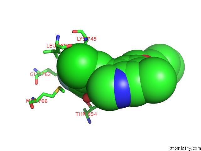
Mono view
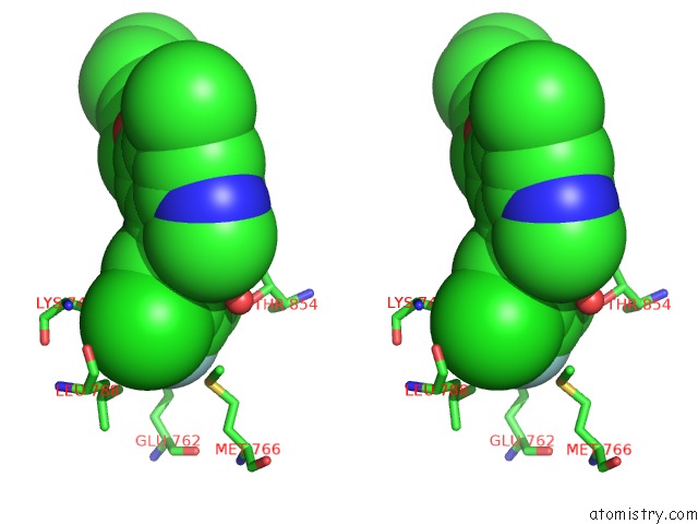
Stereo pair view
Mono view
Stereo pair view
A full contact list of Fluorine with other atoms in the F binding
site number 1 of Crystal Structure of Egfr Kinase Domain L858R Mutation in Complex with Iressa within 5.0Å range:
|
Reference:
C.-H.Yun,
T.J.Boggon,
Y.Li,
S.Woo,
H.Greulich,
M.Meyerson,
M.J.Eck.
Structures of Lung Cancer-Derived Egfr Mutants and Inhibitor Complexes: Mechanism of Activation and Insights Into Differential Inhibitor Sensitivity Cancer Cell V. 11 217 2007.
ISSN: ISSN 1535-6108
PubMed: 17349580
DOI: 10.1016/J.CCR.2006.12.017
Page generated: Wed Jul 31 14:57:04 2024
ISSN: ISSN 1535-6108
PubMed: 17349580
DOI: 10.1016/J.CCR.2006.12.017
Last articles
Zn in 9JPJZn in 9JP7
Zn in 9JPK
Zn in 9JPL
Zn in 9GN6
Zn in 9GN7
Zn in 9GKU
Zn in 9GKW
Zn in 9GKX
Zn in 9GL0