Fluorine »
PDB 2ihk-2jxr »
2jie »
Fluorine in PDB 2jie: Beta-Glucosidase B From Bacillus Polymyxa Complexed with 2- F-Glucose
Enzymatic activity of Beta-Glucosidase B From Bacillus Polymyxa Complexed with 2- F-Glucose
All present enzymatic activity of Beta-Glucosidase B From Bacillus Polymyxa Complexed with 2- F-Glucose:
3.2.1.21;
3.2.1.21;
Protein crystallography data
The structure of Beta-Glucosidase B From Bacillus Polymyxa Complexed with 2- F-Glucose, PDB code: 2jie
was solved by
P.Isorna,
J.Polaina,
J.Sanz-Aparicio,
with X-Ray Crystallography technique. A brief refinement statistics is given in the table below:
| Resolution Low / High (Å) | 25.0 / 2.30 |
| Space group | P 21 21 21 |
| Cell size a, b, c (Å), α, β, γ (°) | 71.236, 74.647, 88.754, 90.00, 90.00, 90.00 |
| R / Rfree (%) | 24.98 / 30.61 |
Fluorine Binding Sites:
The binding sites of Fluorine atom in the Beta-Glucosidase B From Bacillus Polymyxa Complexed with 2- F-Glucose
(pdb code 2jie). This binding sites where shown within
5.0 Angstroms radius around Fluorine atom.
In total only one binding site of Fluorine was determined in the Beta-Glucosidase B From Bacillus Polymyxa Complexed with 2- F-Glucose, PDB code: 2jie:
In total only one binding site of Fluorine was determined in the Beta-Glucosidase B From Bacillus Polymyxa Complexed with 2- F-Glucose, PDB code: 2jie:
Fluorine binding site 1 out of 1 in 2jie
Go back to
Fluorine binding site 1 out
of 1 in the Beta-Glucosidase B From Bacillus Polymyxa Complexed with 2- F-Glucose
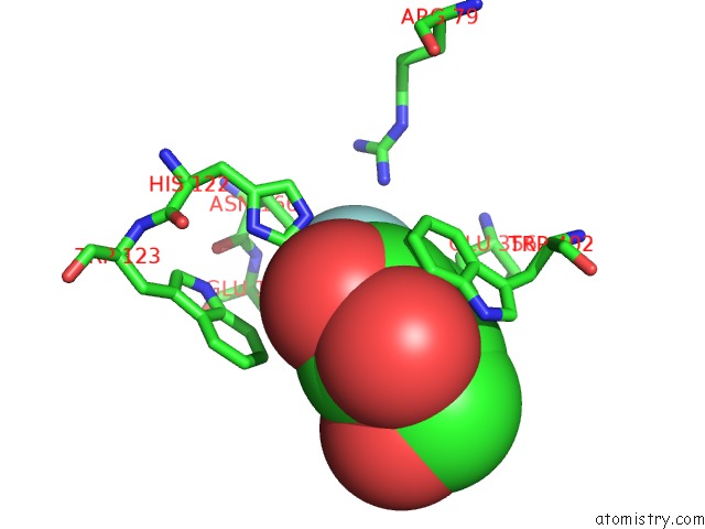
Mono view
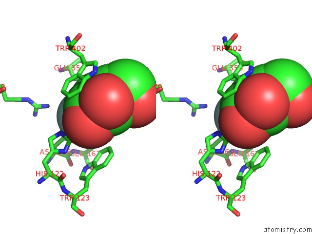
Stereo pair view
Mono view
Stereo pair view
A full contact list of Fluorine with other atoms in the F binding
site number 1 of Beta-Glucosidase B From Bacillus Polymyxa Complexed with 2- F-Glucose within 5.0Å range:
|
Reference:
P.Isorna,
J.Polaina,
L.Latorre-Garcia,
F.J.Canada,
B.Gonzalez,
J.Sanz-Aparicio.
Crystal Structures of Paenibacillus Polymyxa Beta- Glucosidase B Complexes Reveal the Molecular Basis of Substrate Specificity and Give New Insights Into the Catalytic Machinery of Family I Glycosidases. J.Mol.Biol. V. 371 1204 2007.
ISSN: ISSN 0022-2836
PubMed: 17585934
DOI: 10.1016/J.JMB.2007.05.082
Page generated: Wed Jul 31 15:00:26 2024
ISSN: ISSN 0022-2836
PubMed: 17585934
DOI: 10.1016/J.JMB.2007.05.082
Last articles
Zn in 9MJ5Zn in 9HNW
Zn in 9G0L
Zn in 9FNE
Zn in 9DZN
Zn in 9E0I
Zn in 9D32
Zn in 9DAK
Zn in 8ZXC
Zn in 8ZUF