Fluorine »
PDB 2k1q-2ogz »
2k1q »
Fluorine in PDB 2k1q: uc(Nmr) Structure of Hepatitis C Virus NS3 Serine Protease Complexed with the Non-Covalently Bound Phenethylamide Inhibitor
Other elements in 2k1q:
The structure of uc(Nmr) Structure of Hepatitis C Virus NS3 Serine Protease Complexed with the Non-Covalently Bound Phenethylamide Inhibitor also contains other interesting chemical elements:
| Chlorine | (Cl) | 20 atoms |
| Zinc | (Zn) | 20 atoms |
Fluorine Binding Sites:
The binding sites of Fluorine atom in the uc(Nmr) Structure of Hepatitis C Virus NS3 Serine Protease Complexed with the Non-Covalently Bound Phenethylamide Inhibitor
(pdb code 2k1q). This binding sites where shown within
5.0 Angstroms radius around Fluorine atom.
In total 2 binding sites of Fluorine where determined in the uc(Nmr) Structure of Hepatitis C Virus NS3 Serine Protease Complexed with the Non-Covalently Bound Phenethylamide Inhibitor, PDB code: 2k1q:
Jump to Fluorine binding site number: 1; 2;
In total 2 binding sites of Fluorine where determined in the uc(Nmr) Structure of Hepatitis C Virus NS3 Serine Protease Complexed with the Non-Covalently Bound Phenethylamide Inhibitor, PDB code: 2k1q:
Jump to Fluorine binding site number: 1; 2;
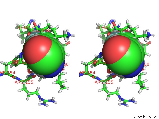
Fluorine binding site 1 out of 2 in 2k1q
Go back to
Fluorine binding site 1 out
of 2 in the uc(Nmr) Structure of Hepatitis C Virus NS3 Serine Protease Complexed with the Non-Covalently Bound Phenethylamide Inhibitor

Mono view

Stereo pair view
Mono view
Stereo pair view
A full contact list of Fluorine with other atoms in the F binding
site number 1 of uc(Nmr) Structure of Hepatitis C Virus NS3 Serine Protease Complexed with the Non-Covalently Bound Phenethylamide Inhibitor within 5.0Å range:
|
Fluorine binding site 2 out of 2 in 2k1q
Go back to
Fluorine binding site 2 out
of 2 in the uc(Nmr) Structure of Hepatitis C Virus NS3 Serine Protease Complexed with the Non-Covalently Bound Phenethylamide Inhibitor

Mono view
Stereo pair view
Mono view
Stereo pair view
A full contact list of Fluorine with other atoms in the F binding
site number 2 of uc(Nmr) Structure of Hepatitis C Virus NS3 Serine Protease Complexed with the Non-Covalently Bound Phenethylamide Inhibitor within 5.0Å range:
|
Reference:
M.Gallo,
M.Pennestri,
M.J.Bottomley,
G.Barbato,
T.Eliseo,
M.Paci,
F.Narjes,
R.De Francesco,
V.Summa,
U.Koch,
R.Bazzo,
D.O.Cicero.
Binding of A Noncovalent Inhibitor Exploiting the S' Region Stabilizes the Hepatitis C Virus NS3 Protease Conformation in the Absence of Cofactor. J.Mol.Biol. V. 385 1142 2009.
ISSN: ISSN 0022-2836
PubMed: 19061898
DOI: 10.1016/J.JMB.2008.11.017
Page generated: Wed Jul 31 15:05:17 2024
ISSN: ISSN 0022-2836
PubMed: 19061898
DOI: 10.1016/J.JMB.2008.11.017
Last articles
Zn in 9MJ5Zn in 9HNW
Zn in 9G0L
Zn in 9FNE
Zn in 9DZN
Zn in 9E0I
Zn in 9D32
Zn in 9DAK
Zn in 8ZXC
Zn in 8ZUF