Fluorine »
PDB 2k1q-2ogz »
2muz »
Fluorine in PDB 2muz: Ssnmr Structure of A Designed Rocker Protein
Fluorine Binding Sites:
The binding sites of Fluorine atom in the Ssnmr Structure of A Designed Rocker Protein
(pdb code 2muz). This binding sites where shown within
5.0 Angstroms radius around Fluorine atom.
In total 8 binding sites of Fluorine where determined in the Ssnmr Structure of A Designed Rocker Protein, PDB code: 2muz:
Jump to Fluorine binding site number: 1; 2; 3; 4; 5; 6; 7; 8;
In total 8 binding sites of Fluorine where determined in the Ssnmr Structure of A Designed Rocker Protein, PDB code: 2muz:
Jump to Fluorine binding site number: 1; 2; 3; 4; 5; 6; 7; 8;
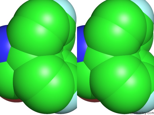
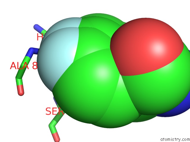
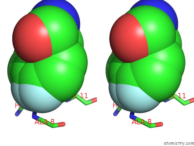
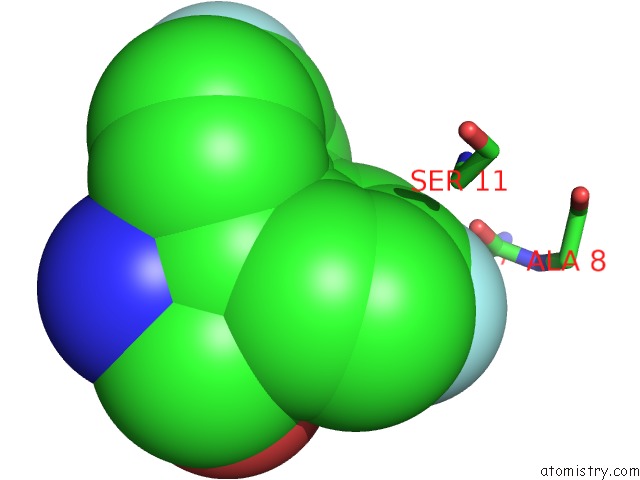
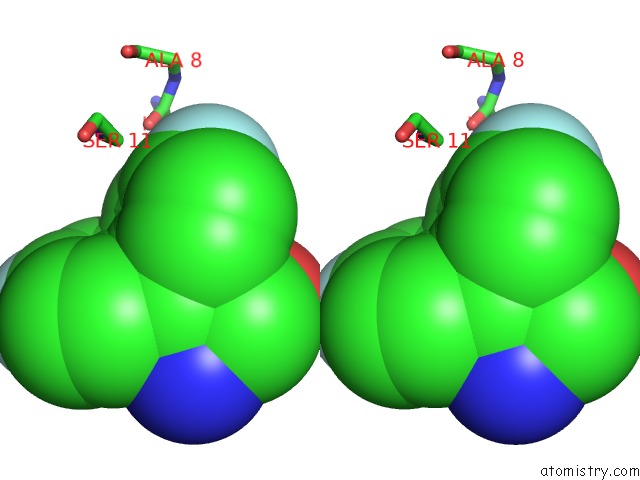
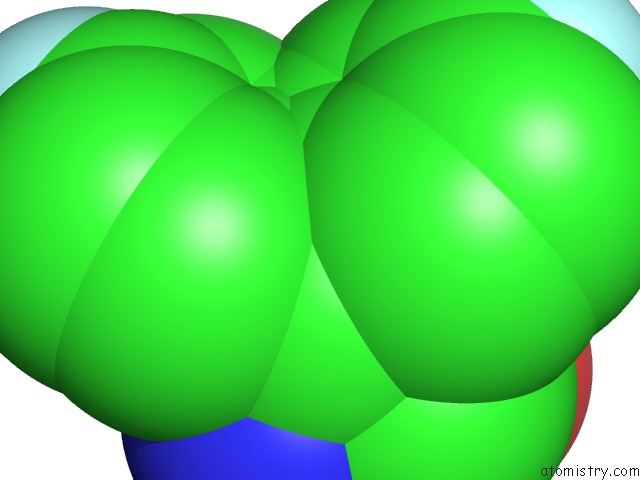
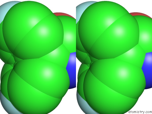
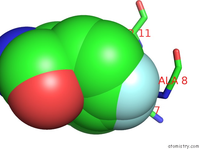
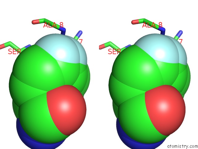
Fluorine binding site 1 out of 8 in 2muz
Go back to
Fluorine binding site 1 out
of 8 in the Ssnmr Structure of A Designed Rocker Protein
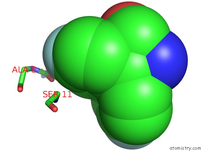
Mono view
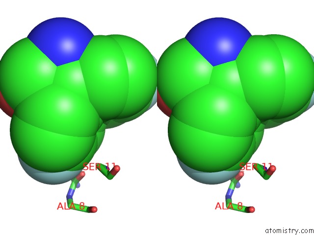
Stereo pair view
Mono view
Stereo pair view
A full contact list of Fluorine with other atoms in the F binding
site number 1 of Ssnmr Structure of A Designed Rocker Protein within 5.0Å range:
|
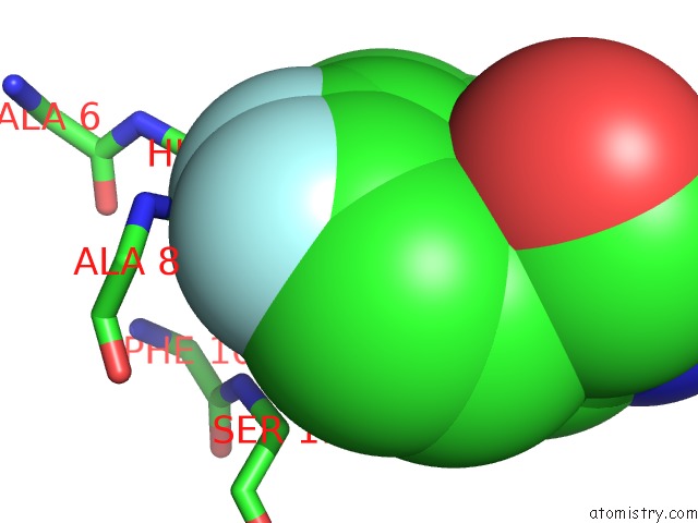
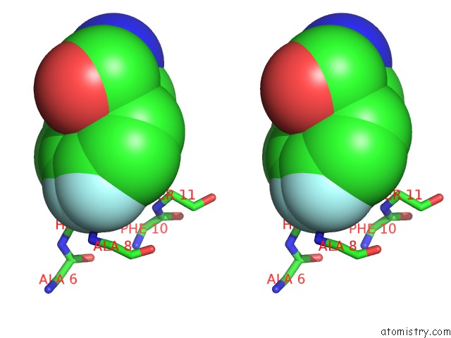
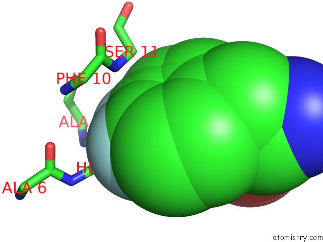
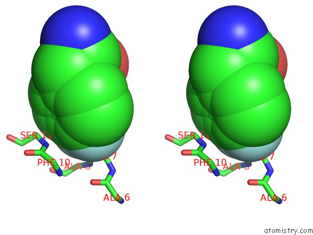
Fluorine binding site 2 out of 8 in 2muz
Go back to
Fluorine binding site 2 out
of 8 in the Ssnmr Structure of A Designed Rocker Protein
Mono view
Stereo pair view
Mono view
Stereo pair view
A full contact list of Fluorine with other atoms in the F binding
site number 2 of Ssnmr Structure of A Designed Rocker Protein within 5.0Å range:
|
Fluorine binding site 3 out of 8 in 2muz
Go back to
Fluorine binding site 3 out
of 8 in the Ssnmr Structure of A Designed Rocker Protein
Mono view
Stereo pair view
Mono view
Stereo pair view
A full contact list of Fluorine with other atoms in the F binding
site number 3 of Ssnmr Structure of A Designed Rocker Protein within 5.0Å range:
|
Fluorine binding site 4 out of 8 in 2muz
Go back to
Fluorine binding site 4 out
of 8 in the Ssnmr Structure of A Designed Rocker Protein
Mono view
Stereo pair view
Mono view
Stereo pair view
A full contact list of Fluorine with other atoms in the F binding
site number 4 of Ssnmr Structure of A Designed Rocker Protein within 5.0Å range:
|
Fluorine binding site 5 out of 8 in 2muz
Go back to
Fluorine binding site 5 out
of 8 in the Ssnmr Structure of A Designed Rocker Protein
Mono view
Stereo pair view
Mono view
Stereo pair view
A full contact list of Fluorine with other atoms in the F binding
site number 5 of Ssnmr Structure of A Designed Rocker Protein within 5.0Å range:
|
Fluorine binding site 6 out of 8 in 2muz
Go back to
Fluorine binding site 6 out
of 8 in the Ssnmr Structure of A Designed Rocker Protein
Mono view
Stereo pair view
Mono view
Stereo pair view
A full contact list of Fluorine with other atoms in the F binding
site number 6 of Ssnmr Structure of A Designed Rocker Protein within 5.0Å range:
|
Fluorine binding site 7 out of 8 in 2muz
Go back to
Fluorine binding site 7 out
of 8 in the Ssnmr Structure of A Designed Rocker Protein
Mono view
Stereo pair view
Mono view
Stereo pair view
A full contact list of Fluorine with other atoms in the F binding
site number 7 of Ssnmr Structure of A Designed Rocker Protein within 5.0Å range:
|
Fluorine binding site 8 out of 8 in 2muz
Go back to
Fluorine binding site 8 out
of 8 in the Ssnmr Structure of A Designed Rocker Protein
Mono view
Stereo pair view
Mono view
Stereo pair view
A full contact list of Fluorine with other atoms in the F binding
site number 8 of Ssnmr Structure of A Designed Rocker Protein within 5.0Å range:
|
Reference:
N.H.Joh,
T.Wang,
M.P.Bhate,
R.Acharya,
Y.Wu,
M.Grabe,
M.Hong,
G.Grigoryan,
W.F.Degrado.
De Novo Design of A Transmembrane Zn(II) Transporting Four-Helix Bundle. Science 2014.
ISSN: ESSN 1095-9203
DOI: 10.1126/SCIENCE.1261172
Page generated: Wed Jul 31 15:07:57 2024
ISSN: ESSN 1095-9203
DOI: 10.1126/SCIENCE.1261172
Last articles
Cl in 7XUDCl in 7XRB
Cl in 7XUC
Cl in 7XRO
Cl in 7XR1
Cl in 7XTF
Cl in 7XQY
Cl in 7XQX
Cl in 7XR0
Cl in 7XQ6