Fluorine »
PDB 2oh4-2pdk »
2opp »
Fluorine in PDB 2opp: Crystal Structure of Hiv-1 Reverse Transcriptase in Complex with GW420867X.
Enzymatic activity of Crystal Structure of Hiv-1 Reverse Transcriptase in Complex with GW420867X.
All present enzymatic activity of Crystal Structure of Hiv-1 Reverse Transcriptase in Complex with GW420867X.:
2.7.7.49;
2.7.7.49;
Protein crystallography data
The structure of Crystal Structure of Hiv-1 Reverse Transcriptase in Complex with GW420867X., PDB code: 2opp
was solved by
J.Ren,
C.E.Nichols,
P.P.Chamberlain,
K.L.Weaver,
S.J.H.Chan,
J.Kleim,
D.K.Stammers,
with X-Ray Crystallography technique. A brief refinement statistics is given in the table below:
| Resolution Low / High (Å) | 29.68 / 2.55 |
| Space group | P 21 21 21 |
| Cell size a, b, c (Å), α, β, γ (°) | 138.500, 115.300, 65.900, 90.00, 90.00, 90.00 |
| R / Rfree (%) | 20.5 / 29.5 |
Other elements in 2opp:
The structure of Crystal Structure of Hiv-1 Reverse Transcriptase in Complex with GW420867X. also contains other interesting chemical elements:
| Magnesium | (Mg) | 1 atom |
Fluorine Binding Sites:
The binding sites of Fluorine atom in the Crystal Structure of Hiv-1 Reverse Transcriptase in Complex with GW420867X.
(pdb code 2opp). This binding sites where shown within
5.0 Angstroms radius around Fluorine atom.
In total only one binding site of Fluorine was determined in the Crystal Structure of Hiv-1 Reverse Transcriptase in Complex with GW420867X., PDB code: 2opp:
In total only one binding site of Fluorine was determined in the Crystal Structure of Hiv-1 Reverse Transcriptase in Complex with GW420867X., PDB code: 2opp:
Fluorine binding site 1 out of 1 in 2opp
Go back to
Fluorine binding site 1 out
of 1 in the Crystal Structure of Hiv-1 Reverse Transcriptase in Complex with GW420867X.
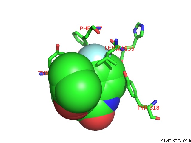
Mono view
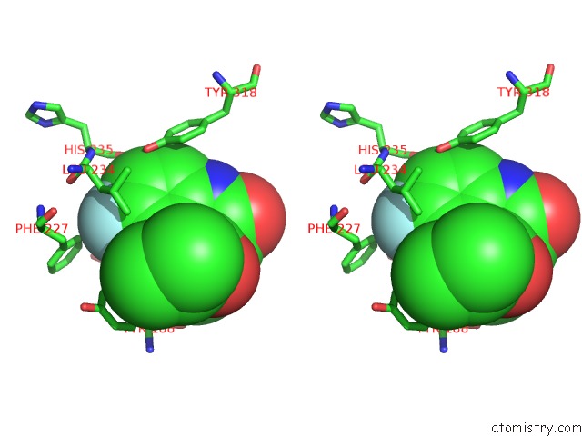
Stereo pair view
Mono view
Stereo pair view
A full contact list of Fluorine with other atoms in the F binding
site number 1 of Crystal Structure of Hiv-1 Reverse Transcriptase in Complex with GW420867X. within 5.0Å range:
|
Reference:
J.Ren,
C.E.Nichols,
P.P.Chamberlain,
K.L.Weaver,
S.A.Short,
J.H.Chan,
J.P.Kleim,
D.K.Stammers.
Relationship of Potency and Resilience to Drug Resistant Mutations For GW420867X Revealed By Crystal Structures of Inhibitor Complexes For Wild-Type, LEU100ILE, LYS101GLU, and TYR188CYS Mutant Hiv-1 Reverse Transcriptases. J.Med.Chem. V. 50 2301 2007.
ISSN: ISSN 0022-2623
PubMed: 17441703
DOI: 10.1021/JM061117M
Page generated: Mon Jul 14 13:53:50 2025
ISSN: ISSN 0022-2623
PubMed: 17441703
DOI: 10.1021/JM061117M
Last articles
Fe in 2YXOFe in 2YRS
Fe in 2YXC
Fe in 2YNM
Fe in 2YVJ
Fe in 2YP1
Fe in 2YU2
Fe in 2YU1
Fe in 2YQB
Fe in 2YOO