Fluorine »
PDB 2oh4-2pdk »
2p4y »
Fluorine in PDB 2p4y: Crystal Structure of Human Ppar-Gamma-Ligand Binding Domain Complexed with An Indole-Based Modulator
Protein crystallography data
The structure of Crystal Structure of Human Ppar-Gamma-Ligand Binding Domain Complexed with An Indole-Based Modulator, PDB code: 2p4y
was solved by
B.M.Mckeever,
with X-Ray Crystallography technique. A brief refinement statistics is given in the table below:
| Resolution Low / High (Å) | 40.34 / 2.25 |
| Space group | C 1 2 1 |
| Cell size a, b, c (Å), α, β, γ (°) | 92.148, 59.859, 118.287, 90.00, 103.76, 90.00 |
| R / Rfree (%) | n/a / n/a |
Other elements in 2p4y:
The structure of Crystal Structure of Human Ppar-Gamma-Ligand Binding Domain Complexed with An Indole-Based Modulator also contains other interesting chemical elements:
| Chlorine | (Cl) | 2 atoms |
Fluorine Binding Sites:
The binding sites of Fluorine atom in the Crystal Structure of Human Ppar-Gamma-Ligand Binding Domain Complexed with An Indole-Based Modulator
(pdb code 2p4y). This binding sites where shown within
5.0 Angstroms radius around Fluorine atom.
In total 6 binding sites of Fluorine where determined in the Crystal Structure of Human Ppar-Gamma-Ligand Binding Domain Complexed with An Indole-Based Modulator, PDB code: 2p4y:
Jump to Fluorine binding site number: 1; 2; 3; 4; 5; 6;
In total 6 binding sites of Fluorine where determined in the Crystal Structure of Human Ppar-Gamma-Ligand Binding Domain Complexed with An Indole-Based Modulator, PDB code: 2p4y:
Jump to Fluorine binding site number: 1; 2; 3; 4; 5; 6;
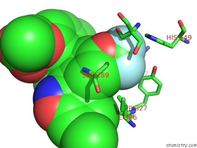
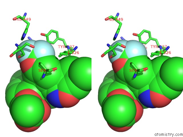
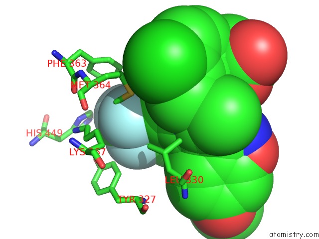
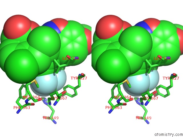
Fluorine binding site 1 out of 6 in 2p4y
Go back to
Fluorine binding site 1 out
of 6 in the Crystal Structure of Human Ppar-Gamma-Ligand Binding Domain Complexed with An Indole-Based Modulator
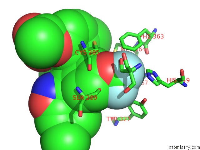
Mono view
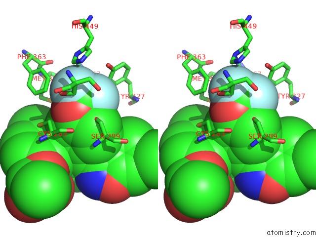
Stereo pair view
Mono view
Stereo pair view
A full contact list of Fluorine with other atoms in the F binding
site number 1 of Crystal Structure of Human Ppar-Gamma-Ligand Binding Domain Complexed with An Indole-Based Modulator within 5.0Å range:
|
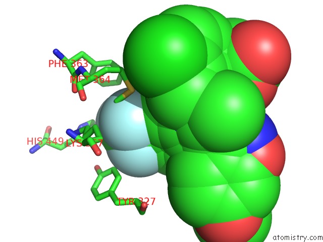
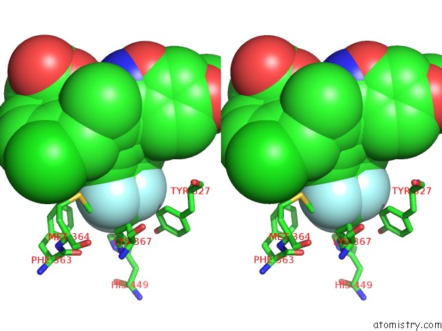
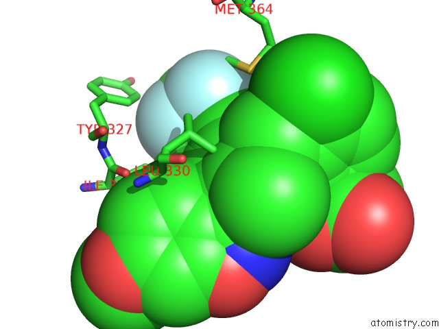
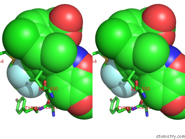
Fluorine binding site 2 out of 6 in 2p4y
Go back to
Fluorine binding site 2 out
of 6 in the Crystal Structure of Human Ppar-Gamma-Ligand Binding Domain Complexed with An Indole-Based Modulator
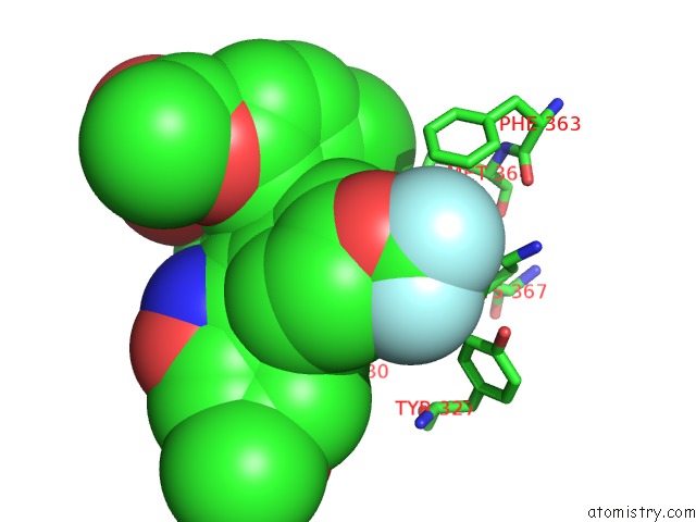
Mono view
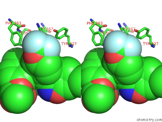
Stereo pair view
Mono view
Stereo pair view
A full contact list of Fluorine with other atoms in the F binding
site number 2 of Crystal Structure of Human Ppar-Gamma-Ligand Binding Domain Complexed with An Indole-Based Modulator within 5.0Å range:
|
Fluorine binding site 3 out of 6 in 2p4y
Go back to
Fluorine binding site 3 out
of 6 in the Crystal Structure of Human Ppar-Gamma-Ligand Binding Domain Complexed with An Indole-Based Modulator
Mono view
Stereo pair view
Mono view
Stereo pair view
A full contact list of Fluorine with other atoms in the F binding
site number 3 of Crystal Structure of Human Ppar-Gamma-Ligand Binding Domain Complexed with An Indole-Based Modulator within 5.0Å range:
|
Fluorine binding site 4 out of 6 in 2p4y
Go back to
Fluorine binding site 4 out
of 6 in the Crystal Structure of Human Ppar-Gamma-Ligand Binding Domain Complexed with An Indole-Based Modulator
Mono view
Stereo pair view
Mono view
Stereo pair view
A full contact list of Fluorine with other atoms in the F binding
site number 4 of Crystal Structure of Human Ppar-Gamma-Ligand Binding Domain Complexed with An Indole-Based Modulator within 5.0Å range:
|
Fluorine binding site 5 out of 6 in 2p4y
Go back to
Fluorine binding site 5 out
of 6 in the Crystal Structure of Human Ppar-Gamma-Ligand Binding Domain Complexed with An Indole-Based Modulator
Mono view
Stereo pair view
Mono view
Stereo pair view
A full contact list of Fluorine with other atoms in the F binding
site number 5 of Crystal Structure of Human Ppar-Gamma-Ligand Binding Domain Complexed with An Indole-Based Modulator within 5.0Å range:
|
Fluorine binding site 6 out of 6 in 2p4y
Go back to
Fluorine binding site 6 out
of 6 in the Crystal Structure of Human Ppar-Gamma-Ligand Binding Domain Complexed with An Indole-Based Modulator
Mono view
Stereo pair view
Mono view
Stereo pair view
A full contact list of Fluorine with other atoms in the F binding
site number 6 of Crystal Structure of Human Ppar-Gamma-Ligand Binding Domain Complexed with An Indole-Based Modulator within 5.0Å range:
|
Reference:
M.Einstein,
T.E.Akiyama,
G.A.Castriota,
C.F.Wang,
B.Mckeever,
R.T.Mosley,
J.W.Becker,
D.E.Moller,
P.T.Meinke,
H.B.Wood,
J.P.Berger.
The Differential Interactions of Peroxisome Proliferator-Activated Receptor Gamma Ligands with TYR473 Is A Physical Basis For Their Unique Biological Activities. Mol.Pharmacol. V. 73 62 2008.
ISSN: ISSN 0026-895X
PubMed: 17940191
DOI: 10.1124/MOL.107.041202
Page generated: Mon Jul 14 13:55:31 2025
ISSN: ISSN 0026-895X
PubMed: 17940191
DOI: 10.1124/MOL.107.041202
Last articles
Fe in 2YXOFe in 2YRS
Fe in 2YXC
Fe in 2YNM
Fe in 2YVJ
Fe in 2YP1
Fe in 2YU2
Fe in 2YU1
Fe in 2YQB
Fe in 2YOO