Fluorine »
PDB 2pdl-2q94 »
2pkk »
Fluorine in PDB 2pkk: Crystal Structure of M Tuberculosis Adenosine Kinase Complexed with 2- Fluro Adenosine
Enzymatic activity of Crystal Structure of M Tuberculosis Adenosine Kinase Complexed with 2- Fluro Adenosine
All present enzymatic activity of Crystal Structure of M Tuberculosis Adenosine Kinase Complexed with 2- Fluro Adenosine:
2.7.1.20;
2.7.1.20;
Protein crystallography data
The structure of Crystal Structure of M Tuberculosis Adenosine Kinase Complexed with 2- Fluro Adenosine, PDB code: 2pkk
was solved by
M.C.M.Reddy,
S.K.Palaninathan,
N.D.Shetty,
J.L.Owen,
M.D.Watson,
J.C.Sacchettini,
Tb Structural Genomics Consortium (Tbsgc),
with X-Ray Crystallography technique. A brief refinement statistics is given in the table below:
| Resolution Low / High (Å) | 62.26 / 1.93 |
| Space group | P 31 2 1 |
| Cell size a, b, c (Å), α, β, γ (°) | 71.916, 71.916, 110.521, 90.00, 90.00, 120.00 |
| R / Rfree (%) | 18.9 / 24 |
Fluorine Binding Sites:
The binding sites of Fluorine atom in the Crystal Structure of M Tuberculosis Adenosine Kinase Complexed with 2- Fluro Adenosine
(pdb code 2pkk). This binding sites where shown within
5.0 Angstroms radius around Fluorine atom.
In total only one binding site of Fluorine was determined in the Crystal Structure of M Tuberculosis Adenosine Kinase Complexed with 2- Fluro Adenosine, PDB code: 2pkk:
In total only one binding site of Fluorine was determined in the Crystal Structure of M Tuberculosis Adenosine Kinase Complexed with 2- Fluro Adenosine, PDB code: 2pkk:
Fluorine binding site 1 out of 1 in 2pkk
Go back to
Fluorine binding site 1 out
of 1 in the Crystal Structure of M Tuberculosis Adenosine Kinase Complexed with 2- Fluro Adenosine
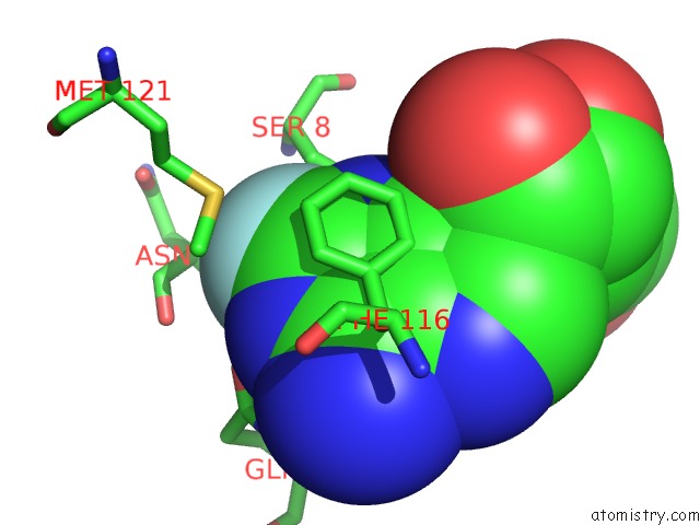
Mono view
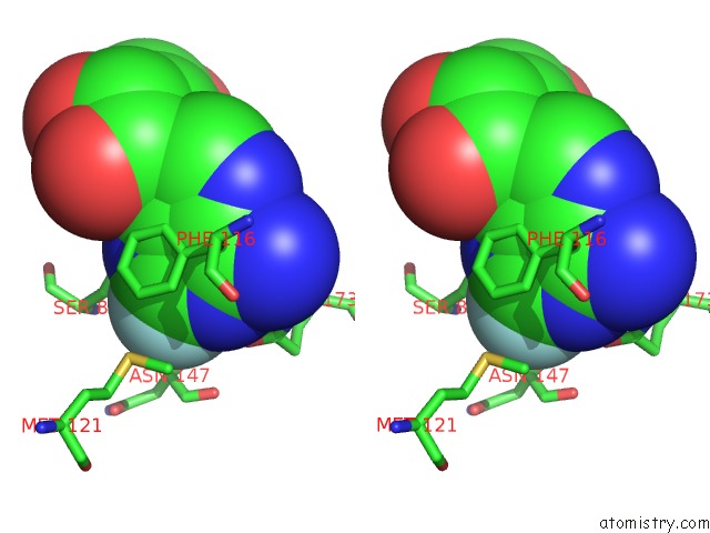
Stereo pair view
Mono view
Stereo pair view
A full contact list of Fluorine with other atoms in the F binding
site number 1 of Crystal Structure of M Tuberculosis Adenosine Kinase Complexed with 2- Fluro Adenosine within 5.0Å range:
|
Reference:
M.C.M.Reddy,
S.K.Palaninathan,
N.D.Shetty,
J.L.Owen,
M.D.Watson,
J.C.Sacchettini.
High Resolution Crystal Structures of Mycobacterium Tuberculosis Adenosine Kinase: Insights Into the Mechanism and Specificity of This Novel Prokaryotic Enzyme J.Biol.Chem. V. 282 27334 2007.
ISSN: ISSN 0021-9258
PubMed: 17597075
DOI: 10.1074/JBC.M703290200
Page generated: Wed Jul 31 15:36:30 2024
ISSN: ISSN 0021-9258
PubMed: 17597075
DOI: 10.1074/JBC.M703290200
Last articles
Zn in 9J0NZn in 9J0O
Zn in 9J0P
Zn in 9FJX
Zn in 9EKB
Zn in 9C0F
Zn in 9CAH
Zn in 9CH0
Zn in 9CH3
Zn in 9CH1