Fluorine »
PDB 2pdl-2q94 »
2prh »
Fluorine in PDB 2prh: The Structures of Apo- and Inhibitor Bound Human Dihydroorotate Dehydrogenase Reveal Conformational Flexibility Within the Inhibitor Binding Site
Enzymatic activity of The Structures of Apo- and Inhibitor Bound Human Dihydroorotate Dehydrogenase Reveal Conformational Flexibility Within the Inhibitor Binding Site
All present enzymatic activity of The Structures of Apo- and Inhibitor Bound Human Dihydroorotate Dehydrogenase Reveal Conformational Flexibility Within the Inhibitor Binding Site:
1.3.99.11;
1.3.99.11;
Protein crystallography data
The structure of The Structures of Apo- and Inhibitor Bound Human Dihydroorotate Dehydrogenase Reveal Conformational Flexibility Within the Inhibitor Binding Site, PDB code: 2prh
was solved by
B.Walse,
V.T.Dufe,
S.Al-Karadaghi,
with X-Ray Crystallography technique. A brief refinement statistics is given in the table below:
| Resolution Low / High (Å) | 19.36 / 2.40 |
| Space group | P 32 2 1 |
| Cell size a, b, c (Å), α, β, γ (°) | 90.550, 90.550, 122.700, 90.00, 90.00, 120.00 |
| R / Rfree (%) | 17.2 / 21.7 |
Other elements in 2prh:
The structure of The Structures of Apo- and Inhibitor Bound Human Dihydroorotate Dehydrogenase Reveal Conformational Flexibility Within the Inhibitor Binding Site also contains other interesting chemical elements:
| Chlorine | (Cl) | 1 atom |
Fluorine Binding Sites:
The binding sites of Fluorine atom in the The Structures of Apo- and Inhibitor Bound Human Dihydroorotate Dehydrogenase Reveal Conformational Flexibility Within the Inhibitor Binding Site
(pdb code 2prh). This binding sites where shown within
5.0 Angstroms radius around Fluorine atom.
In total only one binding site of Fluorine was determined in the The Structures of Apo- and Inhibitor Bound Human Dihydroorotate Dehydrogenase Reveal Conformational Flexibility Within the Inhibitor Binding Site, PDB code: 2prh:
In total only one binding site of Fluorine was determined in the The Structures of Apo- and Inhibitor Bound Human Dihydroorotate Dehydrogenase Reveal Conformational Flexibility Within the Inhibitor Binding Site, PDB code: 2prh:
Fluorine binding site 1 out of 1 in 2prh
Go back to
Fluorine binding site 1 out
of 1 in the The Structures of Apo- and Inhibitor Bound Human Dihydroorotate Dehydrogenase Reveal Conformational Flexibility Within the Inhibitor Binding Site
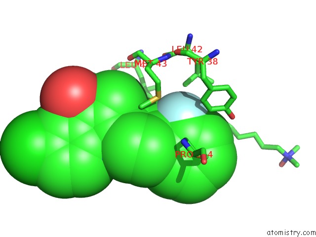
Mono view
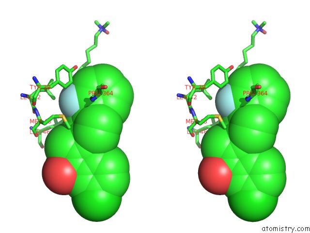
Stereo pair view
Mono view
Stereo pair view
A full contact list of Fluorine with other atoms in the F binding
site number 1 of The Structures of Apo- and Inhibitor Bound Human Dihydroorotate Dehydrogenase Reveal Conformational Flexibility Within the Inhibitor Binding Site within 5.0Å range:
|
Reference:
B.Walse,
V.T.Dufe,
B.Svensson,
I.Fritzson,
L.Dahlberg,
A.Khairoullina,
U.Wellmar,
S.Al-Karadaghi.
The Structures of Human Dihydroorotate Dehydrogenase with and Without Inhibitor Reveal Conformational Flexibility in the Inhibitor and Substrate Binding Sites Biochemistry V. 47 8929 2008.
ISSN: ISSN 0006-2960
PubMed: 18672895
DOI: 10.1021/BI8003318
Page generated: Mon Jul 14 14:04:11 2025
ISSN: ISSN 0006-2960
PubMed: 18672895
DOI: 10.1021/BI8003318
Last articles
Fe in 2YXOFe in 2YRS
Fe in 2YXC
Fe in 2YNM
Fe in 2YVJ
Fe in 2YP1
Fe in 2YU2
Fe in 2YU1
Fe in 2YQB
Fe in 2YOO