Fluorine »
PDB 2pdl-2q94 »
2q94 »
Fluorine in PDB 2q94: E. Coli Methionine Aminopeptidase Mn-Form with Inhibitor A04
Enzymatic activity of E. Coli Methionine Aminopeptidase Mn-Form with Inhibitor A04
All present enzymatic activity of E. Coli Methionine Aminopeptidase Mn-Form with Inhibitor A04:
3.4.11.18;
3.4.11.18;
Protein crystallography data
The structure of E. Coli Methionine Aminopeptidase Mn-Form with Inhibitor A04, PDB code: 2q94
was solved by
Q.-Z.Ye,
with X-Ray Crystallography technique. A brief refinement statistics is given in the table below:
| Resolution Low / High (Å) | 19.91 / 1.63 |
| Space group | P 1 21 1 |
| Cell size a, b, c (Å), α, β, γ (°) | 38.199, 60.605, 50.624, 90.00, 104.80, 90.00 |
| R / Rfree (%) | 21.3 / 23.8 |
Other elements in 2q94:
The structure of E. Coli Methionine Aminopeptidase Mn-Form with Inhibitor A04 also contains other interesting chemical elements:
| Manganese | (Mn) | 2 atoms |
| Sodium | (Na) | 1 atom |
Fluorine Binding Sites:
The binding sites of Fluorine atom in the E. Coli Methionine Aminopeptidase Mn-Form with Inhibitor A04
(pdb code 2q94). This binding sites where shown within
5.0 Angstroms radius around Fluorine atom.
In total 3 binding sites of Fluorine where determined in the E. Coli Methionine Aminopeptidase Mn-Form with Inhibitor A04, PDB code: 2q94:
Jump to Fluorine binding site number: 1; 2; 3;
In total 3 binding sites of Fluorine where determined in the E. Coli Methionine Aminopeptidase Mn-Form with Inhibitor A04, PDB code: 2q94:
Jump to Fluorine binding site number: 1; 2; 3;
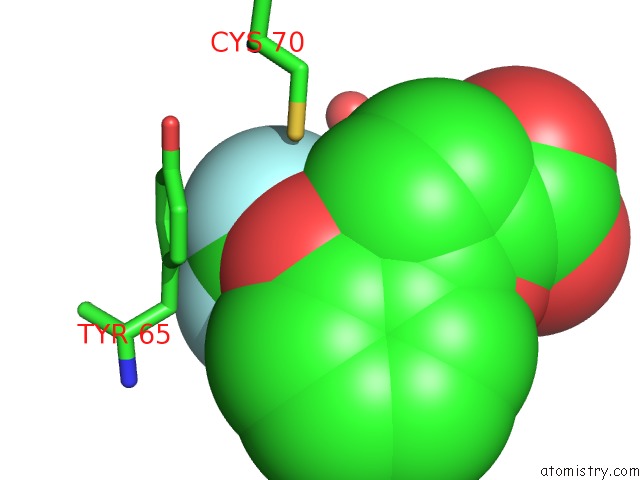
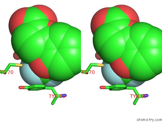
Fluorine binding site 1 out of 3 in 2q94
Go back to
Fluorine binding site 1 out
of 3 in the E. Coli Methionine Aminopeptidase Mn-Form with Inhibitor A04
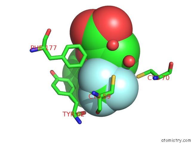
Mono view
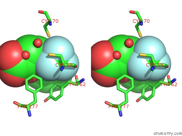
Stereo pair view
Mono view
Stereo pair view
A full contact list of Fluorine with other atoms in the F binding
site number 1 of E. Coli Methionine Aminopeptidase Mn-Form with Inhibitor A04 within 5.0Å range:
|
Fluorine binding site 2 out of 3 in 2q94
Go back to
Fluorine binding site 2 out
of 3 in the E. Coli Methionine Aminopeptidase Mn-Form with Inhibitor A04
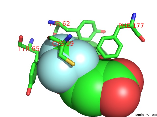
Mono view
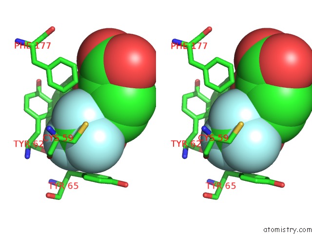
Stereo pair view
Mono view
Stereo pair view
A full contact list of Fluorine with other atoms in the F binding
site number 2 of E. Coli Methionine Aminopeptidase Mn-Form with Inhibitor A04 within 5.0Å range:
|
Fluorine binding site 3 out of 3 in 2q94
Go back to
Fluorine binding site 3 out
of 3 in the E. Coli Methionine Aminopeptidase Mn-Form with Inhibitor A04
Mono view
Stereo pair view
Mono view
Stereo pair view
A full contact list of Fluorine with other atoms in the F binding
site number 3 of E. Coli Methionine Aminopeptidase Mn-Form with Inhibitor A04 within 5.0Å range:
|
Reference:
Z.Q.Ma,
S.X.Xie,
Q.Q.Huang,
F.J.Nan,
T.D.Hurley,
Q.Z.Ye.
Structural Analysis of Inhibition of E. Coli Methionine Aminopeptidase: Implication of Loop Flexibility in Selective Inhibition of Bacterial Enzymes. Bmc Struct.Biol. V. 7 84 2007.
ISSN: ESSN 1472-6807
PubMed: 18093325
DOI: 10.1186/1472-6807-7-84
Page generated: Wed Jul 31 15:45:14 2024
ISSN: ESSN 1472-6807
PubMed: 18093325
DOI: 10.1186/1472-6807-7-84
Last articles
Zn in 9MJ5Zn in 9HNW
Zn in 9G0L
Zn in 9FNE
Zn in 9DZN
Zn in 9E0I
Zn in 9D32
Zn in 9DAK
Zn in 8ZXC
Zn in 8ZUF