Fluorine »
PDB 2q9p-2rbe »
2qfp »
Fluorine in PDB 2qfp: Crystal Structure of Red Kidney Bean Purple Acid Phosphatase in Complex with Fluoride
Enzymatic activity of Crystal Structure of Red Kidney Bean Purple Acid Phosphatase in Complex with Fluoride
All present enzymatic activity of Crystal Structure of Red Kidney Bean Purple Acid Phosphatase in Complex with Fluoride:
3.1.3.2;
3.1.3.2;
Protein crystallography data
The structure of Crystal Structure of Red Kidney Bean Purple Acid Phosphatase in Complex with Fluoride, PDB code: 2qfp
was solved by
L.W.Guddat,
G.S.Schenk,
L.R.Gahan,
T.W.Elliot,
E.Leung,
with X-Ray Crystallography technique. A brief refinement statistics is given in the table below:
| Resolution Low / High (Å) | 33.00 / 2.20 |
| Space group | P 21 21 21 |
| Cell size a, b, c (Å), α, β, γ (°) | 85.719, 188.466, 192.398, 90.00, 90.00, 90.00 |
| R / Rfree (%) | 22.4 / 25.4 |
Other elements in 2qfp:
The structure of Crystal Structure of Red Kidney Bean Purple Acid Phosphatase in Complex with Fluoride also contains other interesting chemical elements:
| Zinc | (Zn) | 4 atoms |
| Iron | (Fe) | 4 atoms |
| Sodium | (Na) | 11 atoms |
Fluorine Binding Sites:
The binding sites of Fluorine atom in the Crystal Structure of Red Kidney Bean Purple Acid Phosphatase in Complex with Fluoride
(pdb code 2qfp). This binding sites where shown within
5.0 Angstroms radius around Fluorine atom.
In total 4 binding sites of Fluorine where determined in the Crystal Structure of Red Kidney Bean Purple Acid Phosphatase in Complex with Fluoride, PDB code: 2qfp:
Jump to Fluorine binding site number: 1; 2; 3; 4;
In total 4 binding sites of Fluorine where determined in the Crystal Structure of Red Kidney Bean Purple Acid Phosphatase in Complex with Fluoride, PDB code: 2qfp:
Jump to Fluorine binding site number: 1; 2; 3; 4;
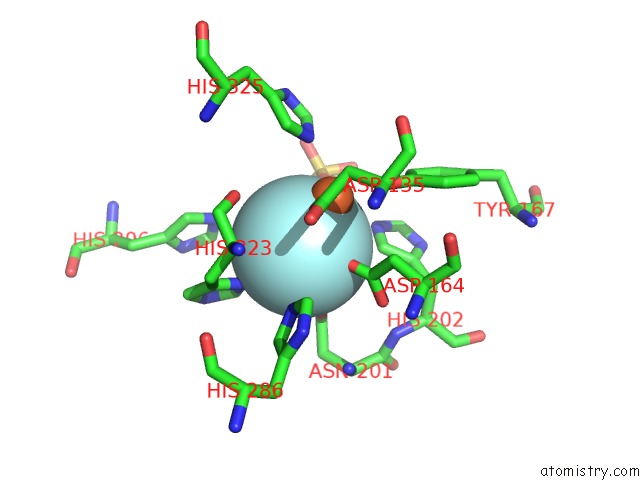
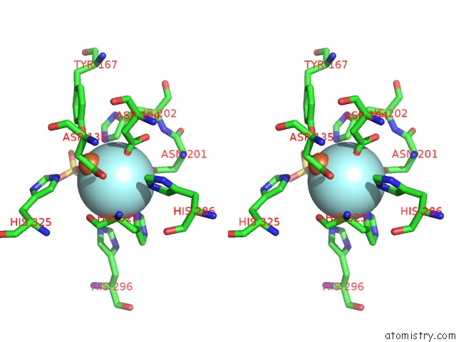
Fluorine binding site 1 out of 4 in 2qfp
Go back to
Fluorine binding site 1 out
of 4 in the Crystal Structure of Red Kidney Bean Purple Acid Phosphatase in Complex with Fluoride
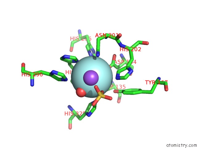
Mono view
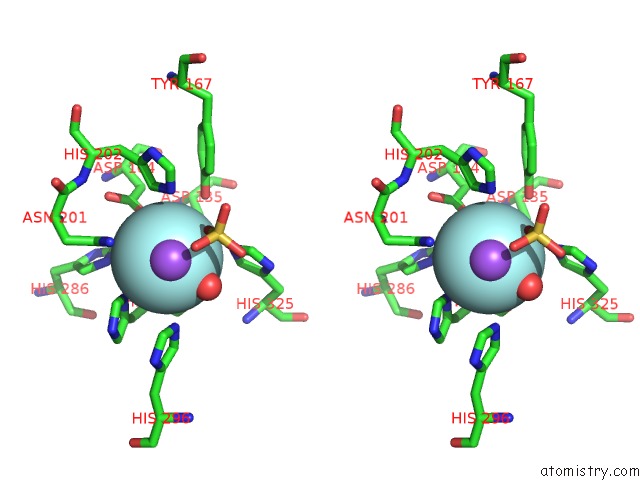
Stereo pair view
Mono view
Stereo pair view
A full contact list of Fluorine with other atoms in the F binding
site number 1 of Crystal Structure of Red Kidney Bean Purple Acid Phosphatase in Complex with Fluoride within 5.0Å range:
|
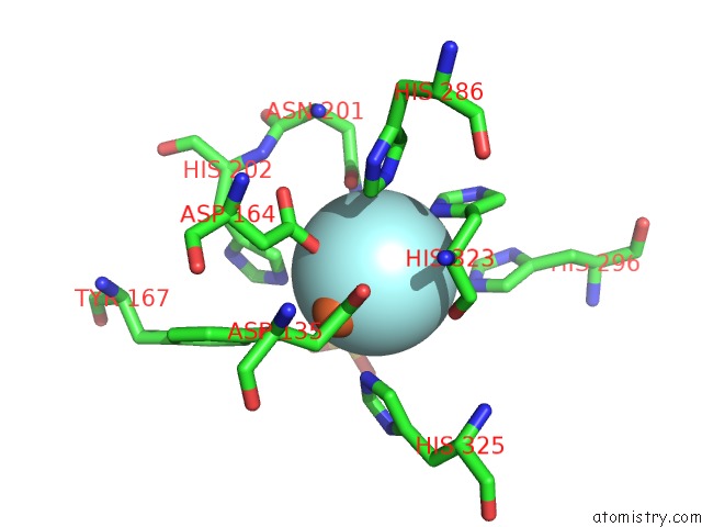
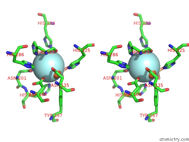
Fluorine binding site 2 out of 4 in 2qfp
Go back to
Fluorine binding site 2 out
of 4 in the Crystal Structure of Red Kidney Bean Purple Acid Phosphatase in Complex with Fluoride
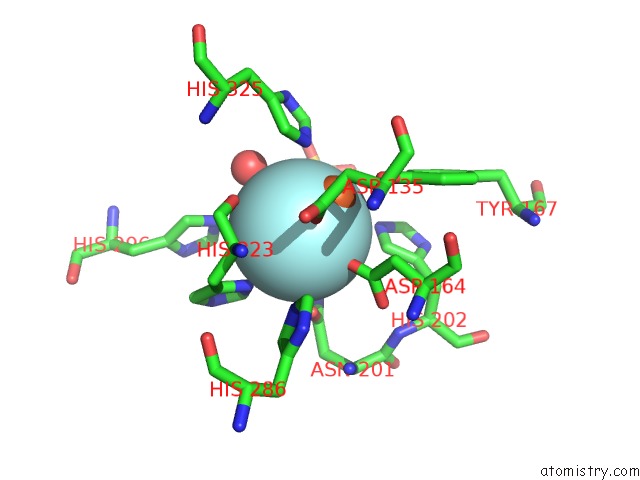
Mono view
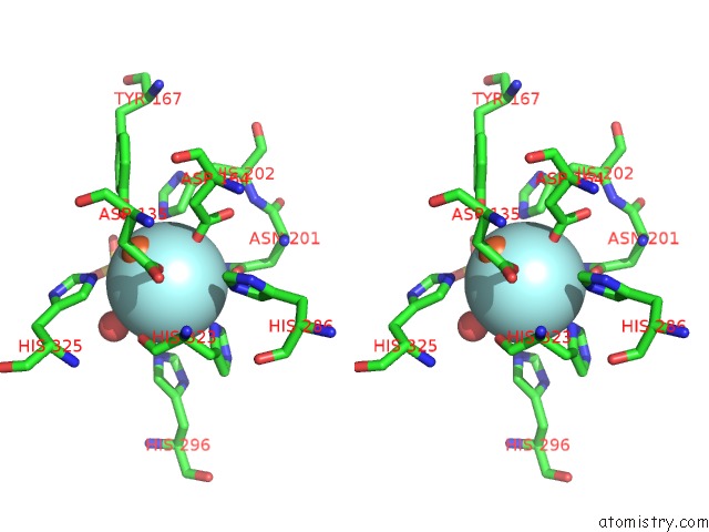
Stereo pair view
Mono view
Stereo pair view
A full contact list of Fluorine with other atoms in the F binding
site number 2 of Crystal Structure of Red Kidney Bean Purple Acid Phosphatase in Complex with Fluoride within 5.0Å range:
|
Fluorine binding site 3 out of 4 in 2qfp
Go back to
Fluorine binding site 3 out
of 4 in the Crystal Structure of Red Kidney Bean Purple Acid Phosphatase in Complex with Fluoride
Mono view
Stereo pair view
Mono view
Stereo pair view
A full contact list of Fluorine with other atoms in the F binding
site number 3 of Crystal Structure of Red Kidney Bean Purple Acid Phosphatase in Complex with Fluoride within 5.0Å range:
|
Fluorine binding site 4 out of 4 in 2qfp
Go back to
Fluorine binding site 4 out
of 4 in the Crystal Structure of Red Kidney Bean Purple Acid Phosphatase in Complex with Fluoride
Mono view
Stereo pair view
Mono view
Stereo pair view
A full contact list of Fluorine with other atoms in the F binding
site number 4 of Crystal Structure of Red Kidney Bean Purple Acid Phosphatase in Complex with Fluoride within 5.0Å range:
|
Reference:
G.Schenk,
T.W.Elliott,
E.Leung,
L.E.Carrington,
N.Mitic,
L.R.Gahan,
L.W.Guddat.
Crystal Structures of A Purple Acid Phosphatase, Representing Different Steps of This Enzyme'S Catalytic Cycle. Bmc Struct.Biol. V. 8 6 2008.
ISSN: ESSN 1472-6807
PubMed: 18234116
DOI: 10.1186/1472-6807-8-6
Page generated: Wed Jul 31 15:47:21 2024
ISSN: ESSN 1472-6807
PubMed: 18234116
DOI: 10.1186/1472-6807-8-6
Last articles
Zn in 9JYWZn in 9IR4
Zn in 9IR3
Zn in 9GMX
Zn in 9GMW
Zn in 9JEJ
Zn in 9ERF
Zn in 9ERE
Zn in 9EGV
Zn in 9EGW