Fluorine »
PDB 2q9p-2rbe »
2qhz »
Fluorine in PDB 2qhz: Crystal Structure of Protease Inhibitor, Mit-1-AC87 in Complex with Wild Type Hiv-1 Protease
Protein crystallography data
The structure of Crystal Structure of Protease Inhibitor, Mit-1-AC87 in Complex with Wild Type Hiv-1 Protease, PDB code: 2qhz
was solved by
C.A.Schiffer,
M.N.L.Nalam,
with X-Ray Crystallography technique. A brief refinement statistics is given in the table below:
| Resolution Low / High (Å) | 42.30 / 1.85 |
| Space group | P 21 21 21 |
| Cell size a, b, c (Å), α, β, γ (°) | 50.737, 58.011, 61.840, 90.00, 90.00, 90.00 |
| R / Rfree (%) | 16.6 / 20.5 |
Fluorine Binding Sites:
The binding sites of Fluorine atom in the Crystal Structure of Protease Inhibitor, Mit-1-AC87 in Complex with Wild Type Hiv-1 Protease
(pdb code 2qhz). This binding sites where shown within
5.0 Angstroms radius around Fluorine atom.
In total 6 binding sites of Fluorine where determined in the Crystal Structure of Protease Inhibitor, Mit-1-AC87 in Complex with Wild Type Hiv-1 Protease, PDB code: 2qhz:
Jump to Fluorine binding site number: 1; 2; 3; 4; 5; 6;
In total 6 binding sites of Fluorine where determined in the Crystal Structure of Protease Inhibitor, Mit-1-AC87 in Complex with Wild Type Hiv-1 Protease, PDB code: 2qhz:
Jump to Fluorine binding site number: 1; 2; 3; 4; 5; 6;
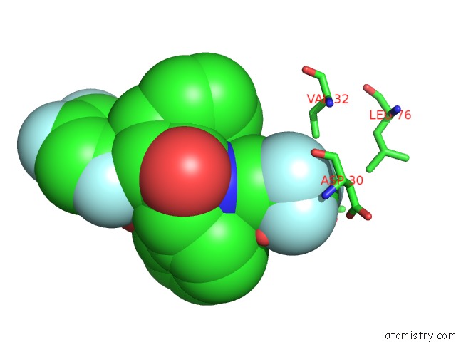
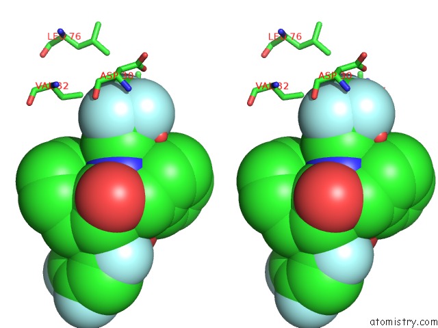
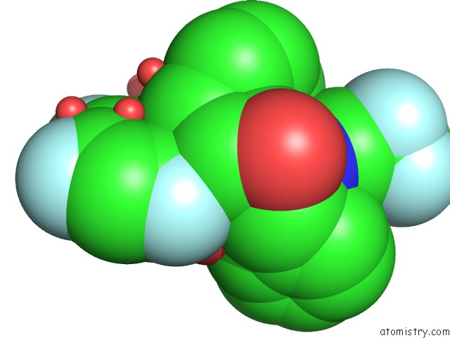
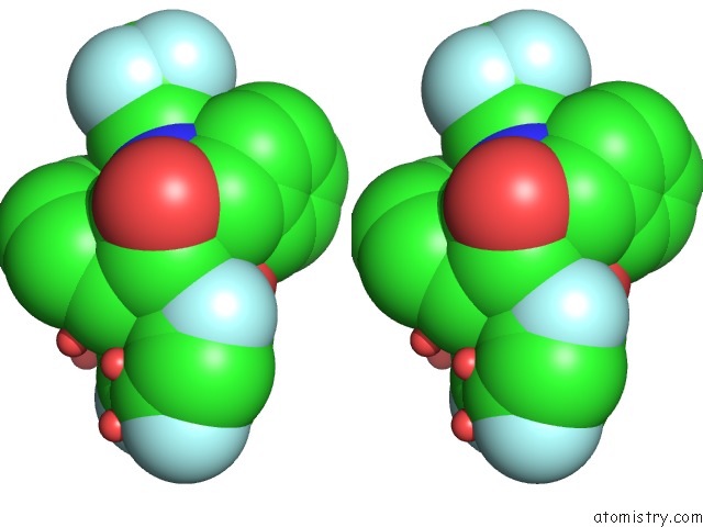
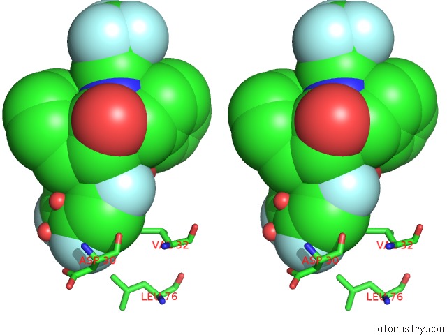
Fluorine binding site 1 out of 6 in 2qhz
Go back to
Fluorine binding site 1 out
of 6 in the Crystal Structure of Protease Inhibitor, Mit-1-AC87 in Complex with Wild Type Hiv-1 Protease

Mono view

Stereo pair view
Mono view
Stereo pair view
A full contact list of Fluorine with other atoms in the F binding
site number 1 of Crystal Structure of Protease Inhibitor, Mit-1-AC87 in Complex with Wild Type Hiv-1 Protease within 5.0Å range:
|
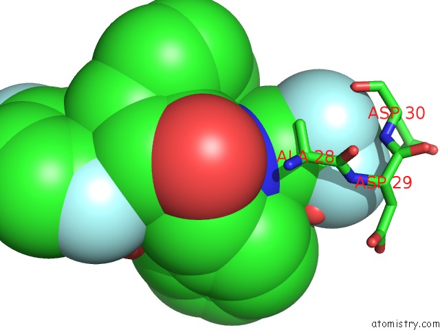
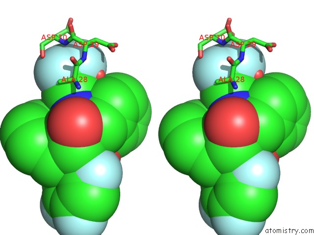
Fluorine binding site 2 out of 6 in 2qhz
Go back to
Fluorine binding site 2 out
of 6 in the Crystal Structure of Protease Inhibitor, Mit-1-AC87 in Complex with Wild Type Hiv-1 Protease
Mono view
Stereo pair view
Mono view
Stereo pair view
A full contact list of Fluorine with other atoms in the F binding
site number 2 of Crystal Structure of Protease Inhibitor, Mit-1-AC87 in Complex with Wild Type Hiv-1 Protease within 5.0Å range:
|
Fluorine binding site 3 out of 6 in 2qhz
Go back to
Fluorine binding site 3 out
of 6 in the Crystal Structure of Protease Inhibitor, Mit-1-AC87 in Complex with Wild Type Hiv-1 Protease
Mono view
Stereo pair view
Mono view
Stereo pair view
A full contact list of Fluorine with other atoms in the F binding
site number 3 of Crystal Structure of Protease Inhibitor, Mit-1-AC87 in Complex with Wild Type Hiv-1 Protease within 5.0Å range:
|
Fluorine binding site 4 out of 6 in 2qhz
Go back to
Fluorine binding site 4 out
of 6 in the Crystal Structure of Protease Inhibitor, Mit-1-AC87 in Complex with Wild Type Hiv-1 Protease
Mono view
Stereo pair view
Mono view
Stereo pair view
A full contact list of Fluorine with other atoms in the F binding
site number 4 of Crystal Structure of Protease Inhibitor, Mit-1-AC87 in Complex with Wild Type Hiv-1 Protease within 5.0Å range:
|
Fluorine binding site 5 out of 6 in 2qhz
Go back to
Fluorine binding site 5 out
of 6 in the Crystal Structure of Protease Inhibitor, Mit-1-AC87 in Complex with Wild Type Hiv-1 Protease

Mono view

Stereo pair view
Mono view
Stereo pair view
A full contact list of Fluorine with other atoms in the F binding
site number 5 of Crystal Structure of Protease Inhibitor, Mit-1-AC87 in Complex with Wild Type Hiv-1 Protease within 5.0Å range:
|
Fluorine binding site 6 out of 6 in 2qhz
Go back to
Fluorine binding site 6 out
of 6 in the Crystal Structure of Protease Inhibitor, Mit-1-AC87 in Complex with Wild Type Hiv-1 Protease

Mono view
Stereo pair view
Mono view
Stereo pair view
A full contact list of Fluorine with other atoms in the F binding
site number 6 of Crystal Structure of Protease Inhibitor, Mit-1-AC87 in Complex with Wild Type Hiv-1 Protease within 5.0Å range:
|
Reference:
M.D.Altman,
A.Ali,
G.S.Reddy,
M.N.Nalam,
S.G.Anjum,
H.Cao,
S.Chellappan,
V.Kairys,
M.X.Fernandes,
M.K.Gilson,
C.A.Schiffer,
T.M.Rana,
B.Tidor.
Hiv-1 Protease Inhibitors From Inverse Design in the Substrate Envelope Exhibit Subnanomolar Binding to Drug-Resistant Variants. J.Am.Chem.Soc. V. 130 6099 2008.
ISSN: ISSN 0002-7863
PubMed: 18412349
DOI: 10.1021/JA076558P
Page generated: Wed Jul 31 15:47:21 2024
ISSN: ISSN 0002-7863
PubMed: 18412349
DOI: 10.1021/JA076558P
Last articles
Zn in 9JYWZn in 9IR4
Zn in 9IR3
Zn in 9GMX
Zn in 9GMW
Zn in 9JEJ
Zn in 9ERF
Zn in 9ERE
Zn in 9EGV
Zn in 9EGW