Fluorine »
PDB 2rfn-2vfz »
2rhw »
Fluorine in PDB 2rhw: Crystal Structure of the S112A Mutant of A C-C Hydrolase, Bphd From Burkholderia Xenovorans LB400, in Complex with 3,10-Di-Fluoro Hopda
Protein crystallography data
The structure of Crystal Structure of the S112A Mutant of A C-C Hydrolase, Bphd From Burkholderia Xenovorans LB400, in Complex with 3,10-Di-Fluoro Hopda, PDB code: 2rhw
was solved by
S.Bhowmik,
J.T.Bolin,
with X-Ray Crystallography technique. A brief refinement statistics is given in the table below:
| Resolution Low / High (Å) | 27.80 / 1.57 |
| Space group | I 41 2 2 |
| Cell size a, b, c (Å), α, β, γ (°) | 117.956, 117.956, 87.193, 90.00, 90.00, 90.00 |
| R / Rfree (%) | 17.1 / 20 |
Other elements in 2rhw:
The structure of Crystal Structure of the S112A Mutant of A C-C Hydrolase, Bphd From Burkholderia Xenovorans LB400, in Complex with 3,10-Di-Fluoro Hopda also contains other interesting chemical elements:
| Sodium | (Na) | 1 atom |
Fluorine Binding Sites:
The binding sites of Fluorine atom in the Crystal Structure of the S112A Mutant of A C-C Hydrolase, Bphd From Burkholderia Xenovorans LB400, in Complex with 3,10-Di-Fluoro Hopda
(pdb code 2rhw). This binding sites where shown within
5.0 Angstroms radius around Fluorine atom.
In total 2 binding sites of Fluorine where determined in the Crystal Structure of the S112A Mutant of A C-C Hydrolase, Bphd From Burkholderia Xenovorans LB400, in Complex with 3,10-Di-Fluoro Hopda, PDB code: 2rhw:
Jump to Fluorine binding site number: 1; 2;
In total 2 binding sites of Fluorine where determined in the Crystal Structure of the S112A Mutant of A C-C Hydrolase, Bphd From Burkholderia Xenovorans LB400, in Complex with 3,10-Di-Fluoro Hopda, PDB code: 2rhw:
Jump to Fluorine binding site number: 1; 2;
Fluorine binding site 1 out of 2 in 2rhw
Go back to
Fluorine binding site 1 out
of 2 in the Crystal Structure of the S112A Mutant of A C-C Hydrolase, Bphd From Burkholderia Xenovorans LB400, in Complex with 3,10-Di-Fluoro Hopda
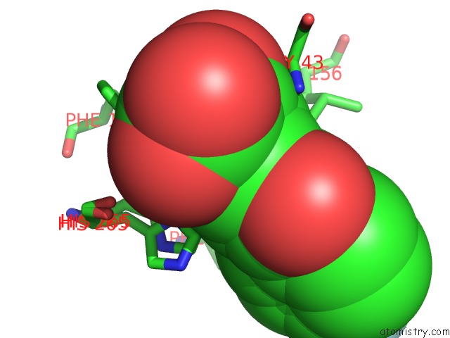
Mono view
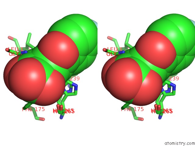
Stereo pair view
Mono view
Stereo pair view
A full contact list of Fluorine with other atoms in the F binding
site number 1 of Crystal Structure of the S112A Mutant of A C-C Hydrolase, Bphd From Burkholderia Xenovorans LB400, in Complex with 3,10-Di-Fluoro Hopda within 5.0Å range:
|
Fluorine binding site 2 out of 2 in 2rhw
Go back to
Fluorine binding site 2 out
of 2 in the Crystal Structure of the S112A Mutant of A C-C Hydrolase, Bphd From Burkholderia Xenovorans LB400, in Complex with 3,10-Di-Fluoro Hopda
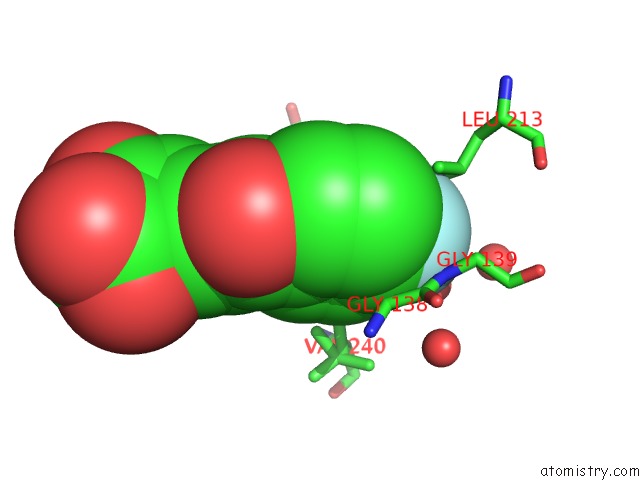
Mono view
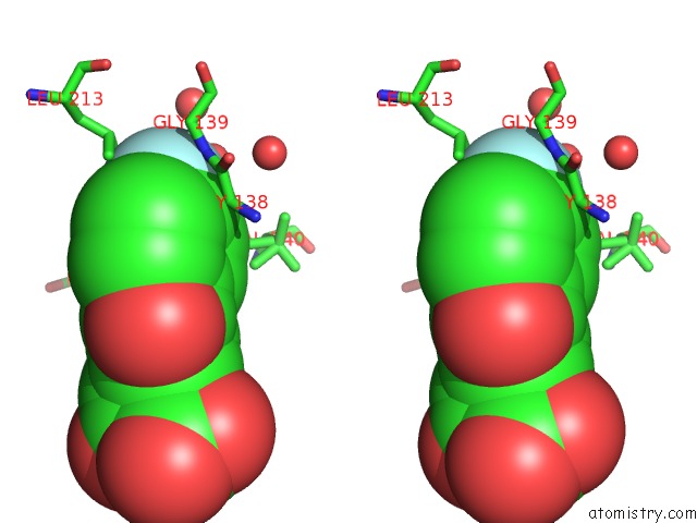
Stereo pair view
Mono view
Stereo pair view
A full contact list of Fluorine with other atoms in the F binding
site number 2 of Crystal Structure of the S112A Mutant of A C-C Hydrolase, Bphd From Burkholderia Xenovorans LB400, in Complex with 3,10-Di-Fluoro Hopda within 5.0Å range:
|
Reference:
S.Bhowmik,
G.P.Horsman,
J.T.Bolin,
L.D.Eltis.
The Molecular Basis For Inhibition of Bphd, A C-C Bond Hydrolase Involved in Polychlorinated Biphenyls Degradation: Large 3-Substituents Prevent Tautomerization. J.Biol.Chem. V. 282 36377 2007.
ISSN: ISSN 0021-9258
PubMed: 17932031
DOI: 10.1074/JBC.M707035200
Page generated: Wed Jul 31 16:00:35 2024
ISSN: ISSN 0021-9258
PubMed: 17932031
DOI: 10.1074/JBC.M707035200
Last articles
Zn in 9MJ5Zn in 9HNW
Zn in 9G0L
Zn in 9FNE
Zn in 9DZN
Zn in 9E0I
Zn in 9D32
Zn in 9DAK
Zn in 8ZXC
Zn in 8ZUF