Fluorine »
PDB 2rfn-2vfz »
2v7v »
Fluorine in PDB 2v7v: X-Ray Crystal Structure of 5'-Fluorodeoxyadenosine Synthase From Streptomyces Cattleya Complexed with 5'-Fluorodeoxyadenosine
Enzymatic activity of X-Ray Crystal Structure of 5'-Fluorodeoxyadenosine Synthase From Streptomyces Cattleya Complexed with 5'-Fluorodeoxyadenosine
All present enzymatic activity of X-Ray Crystal Structure of 5'-Fluorodeoxyadenosine Synthase From Streptomyces Cattleya Complexed with 5'-Fluorodeoxyadenosine:
2.5.1.63;
2.5.1.63;
Protein crystallography data
The structure of X-Ray Crystal Structure of 5'-Fluorodeoxyadenosine Synthase From Streptomyces Cattleya Complexed with 5'-Fluorodeoxyadenosine, PDB code: 2v7v
was solved by
X.Zhu,
D.O'hagan,
J.H.Naismith,
with X-Ray Crystallography technique. A brief refinement statistics is given in the table below:
| Resolution Low / High (Å) | 37.16 / 1.94 |
| Space group | C 2 2 21 |
| Cell size a, b, c (Å), α, β, γ (°) | 75.892, 129.946, 184.491, 90.00, 90.00, 90.00 |
| R / Rfree (%) | 18 / 22.1 |
Fluorine Binding Sites:
The binding sites of Fluorine atom in the X-Ray Crystal Structure of 5'-Fluorodeoxyadenosine Synthase From Streptomyces Cattleya Complexed with 5'-Fluorodeoxyadenosine
(pdb code 2v7v). This binding sites where shown within
5.0 Angstroms radius around Fluorine atom.
In total 3 binding sites of Fluorine where determined in the X-Ray Crystal Structure of 5'-Fluorodeoxyadenosine Synthase From Streptomyces Cattleya Complexed with 5'-Fluorodeoxyadenosine, PDB code: 2v7v:
Jump to Fluorine binding site number: 1; 2; 3;
In total 3 binding sites of Fluorine where determined in the X-Ray Crystal Structure of 5'-Fluorodeoxyadenosine Synthase From Streptomyces Cattleya Complexed with 5'-Fluorodeoxyadenosine, PDB code: 2v7v:
Jump to Fluorine binding site number: 1; 2; 3;
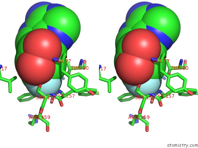
Fluorine binding site 1 out of 3 in 2v7v
Go back to
Fluorine binding site 1 out
of 3 in the X-Ray Crystal Structure of 5'-Fluorodeoxyadenosine Synthase From Streptomyces Cattleya Complexed with 5'-Fluorodeoxyadenosine
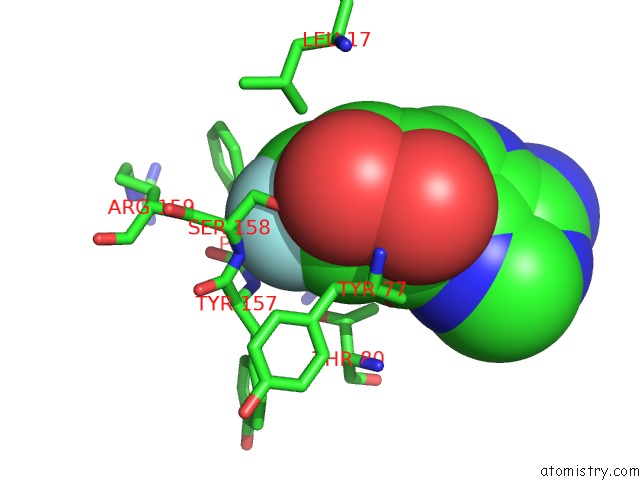
Mono view
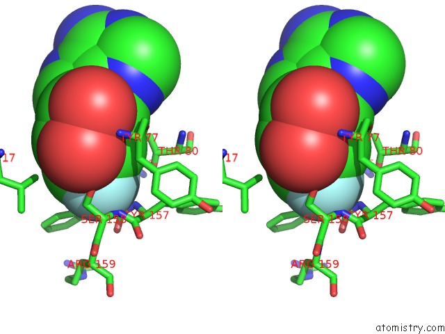
Stereo pair view
Mono view
Stereo pair view
A full contact list of Fluorine with other atoms in the F binding
site number 1 of X-Ray Crystal Structure of 5'-Fluorodeoxyadenosine Synthase From Streptomyces Cattleya Complexed with 5'-Fluorodeoxyadenosine within 5.0Å range:
|
Fluorine binding site 2 out of 3 in 2v7v
Go back to
Fluorine binding site 2 out
of 3 in the X-Ray Crystal Structure of 5'-Fluorodeoxyadenosine Synthase From Streptomyces Cattleya Complexed with 5'-Fluorodeoxyadenosine
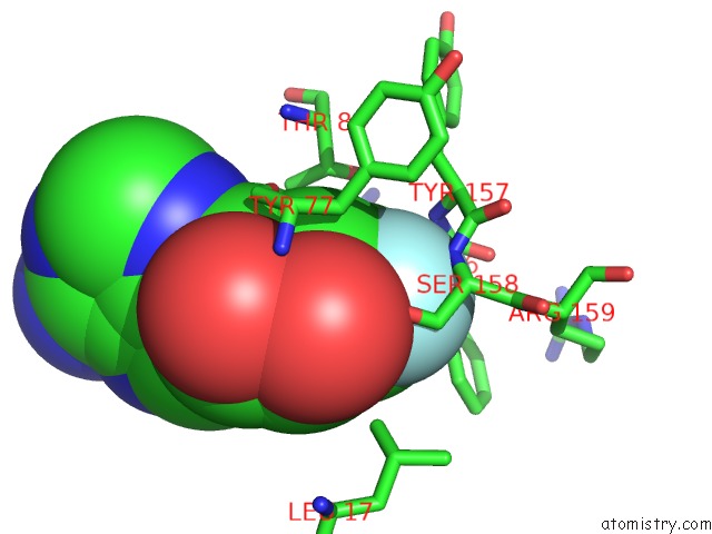
Mono view

Stereo pair view
Mono view
Stereo pair view
A full contact list of Fluorine with other atoms in the F binding
site number 2 of X-Ray Crystal Structure of 5'-Fluorodeoxyadenosine Synthase From Streptomyces Cattleya Complexed with 5'-Fluorodeoxyadenosine within 5.0Å range:
|
Fluorine binding site 3 out of 3 in 2v7v
Go back to
Fluorine binding site 3 out
of 3 in the X-Ray Crystal Structure of 5'-Fluorodeoxyadenosine Synthase From Streptomyces Cattleya Complexed with 5'-Fluorodeoxyadenosine
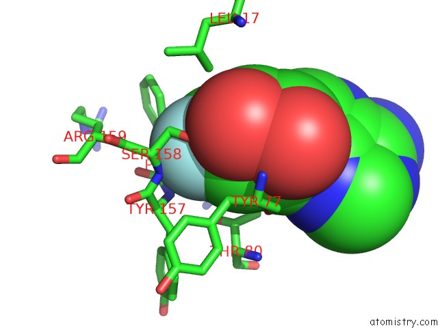
Mono view
Stereo pair view
Mono view
Stereo pair view
A full contact list of Fluorine with other atoms in the F binding
site number 3 of X-Ray Crystal Structure of 5'-Fluorodeoxyadenosine Synthase From Streptomyces Cattleya Complexed with 5'-Fluorodeoxyadenosine within 5.0Å range:
|
Reference:
X.Zhu,
D.A.Robinson,
A.R.Mcewan,
D.O'hagan,
J.H.Naismith.
Mechanism of Enzymatic Fluorination in Streptomyces Cattleya. J. Am. Chem. Soc. V. 129 14597 2007.
ISSN: ESSN 1520-5126
PubMed: 17985882
DOI: 10.1021/JA0731569
Page generated: Wed Jul 31 16:03:20 2024
ISSN: ESSN 1520-5126
PubMed: 17985882
DOI: 10.1021/JA0731569
Last articles
Zn in 9J0NZn in 9J0O
Zn in 9J0P
Zn in 9FJX
Zn in 9EKB
Zn in 9C0F
Zn in 9CAH
Zn in 9CH0
Zn in 9CH3
Zn in 9CH1