Fluorine »
PDB 2rfn-2vfz »
2vas »
Fluorine in PDB 2vas: Myosin VI (Md-INSERT2-Cam, Delta-INSERT1) Post-Rigor State
Protein crystallography data
The structure of Myosin VI (Md-INSERT2-Cam, Delta-INSERT1) Post-Rigor State, PDB code: 2vas
was solved by
J.Menetrey,
P.Llinas,
J.Cicolari,
G.Squires,
X.Liu,
A.Li,
H.L.Sweeney,
A.Houdusse,
with X-Ray Crystallography technique. A brief refinement statistics is given in the table below:
| Resolution Low / High (Å) | 48.28 / 2.40 |
| Space group | P 21 21 21 |
| Cell size a, b, c (Å), α, β, γ (°) | 73.420, 100.470, 174.070, 90.00, 90.00, 90.00 |
| R / Rfree (%) | 21.7 / 25.6 |
Other elements in 2vas:
The structure of Myosin VI (Md-INSERT2-Cam, Delta-INSERT1) Post-Rigor State also contains other interesting chemical elements:
| Magnesium | (Mg) | 1 atom |
| Calcium | (Ca) | 4 atoms |
Fluorine Binding Sites:
The binding sites of Fluorine atom in the Myosin VI (Md-INSERT2-Cam, Delta-INSERT1) Post-Rigor State
(pdb code 2vas). This binding sites where shown within
5.0 Angstroms radius around Fluorine atom.
In total 3 binding sites of Fluorine where determined in the Myosin VI (Md-INSERT2-Cam, Delta-INSERT1) Post-Rigor State, PDB code: 2vas:
Jump to Fluorine binding site number: 1; 2; 3;
In total 3 binding sites of Fluorine where determined in the Myosin VI (Md-INSERT2-Cam, Delta-INSERT1) Post-Rigor State, PDB code: 2vas:
Jump to Fluorine binding site number: 1; 2; 3;
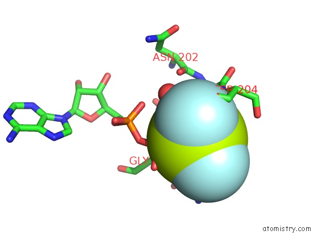
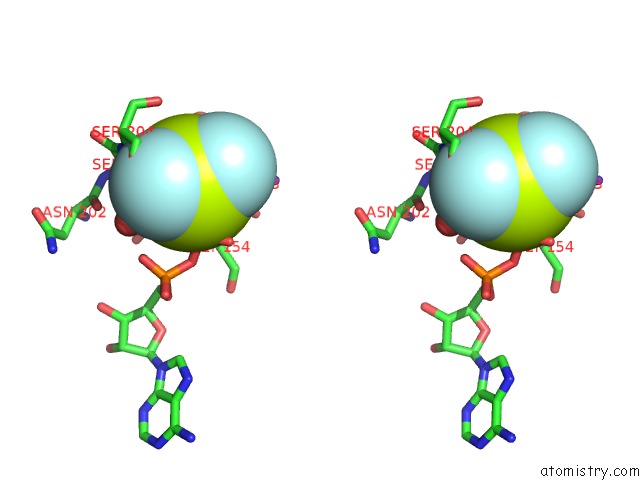
Fluorine binding site 1 out of 3 in 2vas
Go back to
Fluorine binding site 1 out
of 3 in the Myosin VI (Md-INSERT2-Cam, Delta-INSERT1) Post-Rigor State
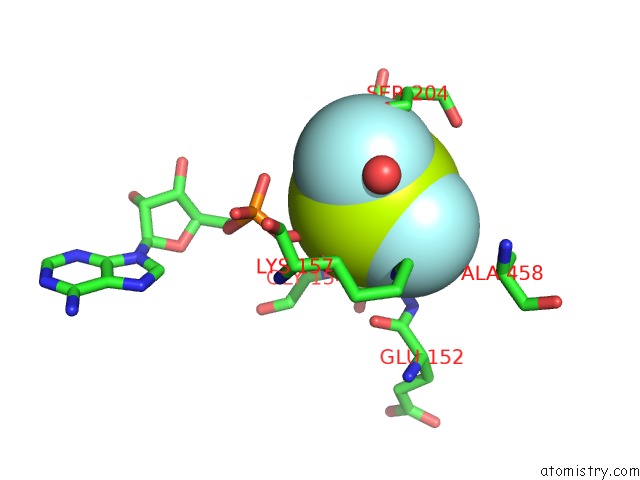
Mono view
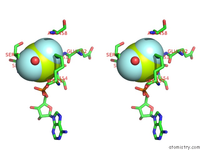
Stereo pair view
Mono view
Stereo pair view
A full contact list of Fluorine with other atoms in the F binding
site number 1 of Myosin VI (Md-INSERT2-Cam, Delta-INSERT1) Post-Rigor State within 5.0Å range:
|
Fluorine binding site 2 out of 3 in 2vas
Go back to
Fluorine binding site 2 out
of 3 in the Myosin VI (Md-INSERT2-Cam, Delta-INSERT1) Post-Rigor State
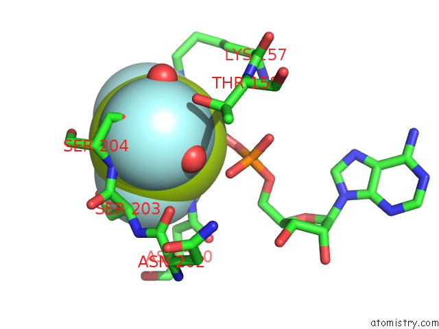
Mono view
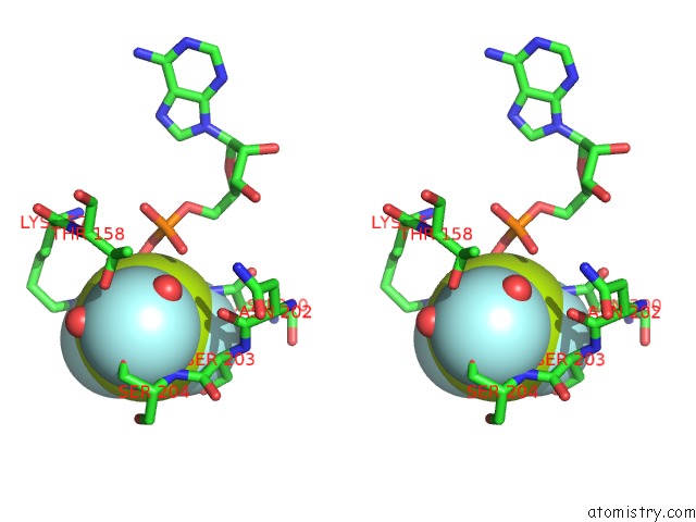
Stereo pair view
Mono view
Stereo pair view
A full contact list of Fluorine with other atoms in the F binding
site number 2 of Myosin VI (Md-INSERT2-Cam, Delta-INSERT1) Post-Rigor State within 5.0Å range:
|
Fluorine binding site 3 out of 3 in 2vas
Go back to
Fluorine binding site 3 out
of 3 in the Myosin VI (Md-INSERT2-Cam, Delta-INSERT1) Post-Rigor State
Mono view
Stereo pair view
Mono view
Stereo pair view
A full contact list of Fluorine with other atoms in the F binding
site number 3 of Myosin VI (Md-INSERT2-Cam, Delta-INSERT1) Post-Rigor State within 5.0Å range:
|
Reference:
J.Menetrey,
P.Llinas,
J.Cicolari,
G.Squires,
X.Liu,
A.Li,
H.L.Sweeney,
A.Houdusse.
The Post-Rigor Structure of Myosin VI and Implications For the Recovery Stroke. Embo J. V. 27 244 2008.
ISSN: ISSN 0261-4189
PubMed: 18046460
DOI: 10.1038/SJ.EMBOJ.7601937
Page generated: Wed Jul 31 16:06:55 2024
ISSN: ISSN 0261-4189
PubMed: 18046460
DOI: 10.1038/SJ.EMBOJ.7601937
Last articles
Zn in 9J0NZn in 9J0O
Zn in 9J0P
Zn in 9FJX
Zn in 9EKB
Zn in 9C0F
Zn in 9CAH
Zn in 9CH0
Zn in 9CH3
Zn in 9CH1