Fluorine »
PDB 2vh0-2wbk »
2vui »
Fluorine in PDB 2vui: Crystal Structure of the Hupr Receiver Domain in Inhibitory Phospho-State
Protein crystallography data
The structure of Crystal Structure of the Hupr Receiver Domain in Inhibitory Phospho-State, PDB code: 2vui
was solved by
K.M.Davies,
E.D.Lowe,
C.Venien-Bryan,
L.N.Johnson,
with X-Ray Crystallography technique. A brief refinement statistics is given in the table below:
| Resolution Low / High (Å) | 23.30 / 2.90 |
| Space group | P 32 1 2 |
| Cell size a, b, c (Å), α, β, γ (°) | 81.893, 81.893, 61.812, 90.00, 90.00, 120.00 |
| R / Rfree (%) | 25.78 / 32.02 |
Other elements in 2vui:
The structure of Crystal Structure of the Hupr Receiver Domain in Inhibitory Phospho-State also contains other interesting chemical elements:
| Magnesium | (Mg) | 2 atoms |
Fluorine Binding Sites:
The binding sites of Fluorine atom in the Crystal Structure of the Hupr Receiver Domain in Inhibitory Phospho-State
(pdb code 2vui). This binding sites where shown within
5.0 Angstroms radius around Fluorine atom.
In total 3 binding sites of Fluorine where determined in the Crystal Structure of the Hupr Receiver Domain in Inhibitory Phospho-State, PDB code: 2vui:
Jump to Fluorine binding site number: 1; 2; 3;
In total 3 binding sites of Fluorine where determined in the Crystal Structure of the Hupr Receiver Domain in Inhibitory Phospho-State, PDB code: 2vui:
Jump to Fluorine binding site number: 1; 2; 3;
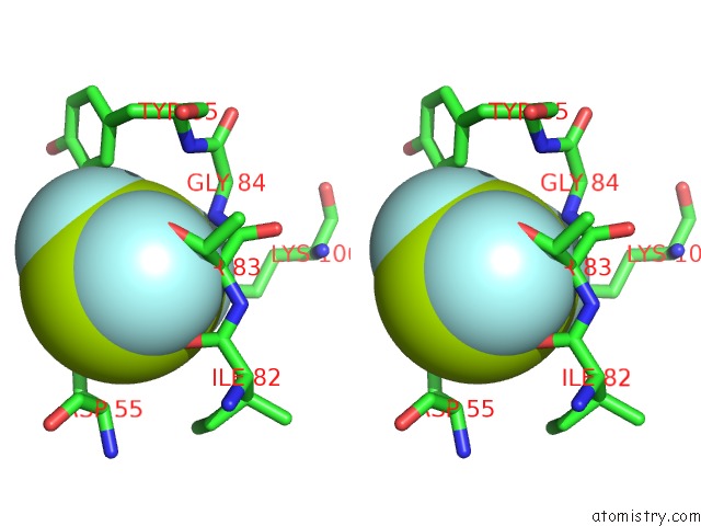
Fluorine binding site 1 out of 3 in 2vui
Go back to
Fluorine binding site 1 out
of 3 in the Crystal Structure of the Hupr Receiver Domain in Inhibitory Phospho-State
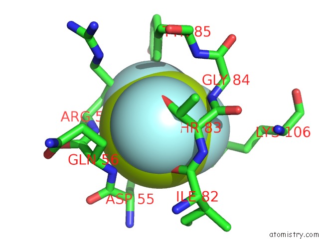
Mono view
Stereo pair view
Mono view
Stereo pair view
A full contact list of Fluorine with other atoms in the F binding
site number 1 of Crystal Structure of the Hupr Receiver Domain in Inhibitory Phospho-State within 5.0Å range:
|
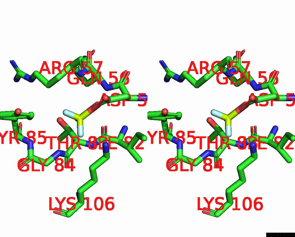
Fluorine binding site 2 out of 3 in 2vui
Go back to
Fluorine binding site 2 out
of 3 in the Crystal Structure of the Hupr Receiver Domain in Inhibitory Phospho-State

Mono view
Stereo pair view
Mono view
Stereo pair view
A full contact list of Fluorine with other atoms in the F binding
site number 2 of Crystal Structure of the Hupr Receiver Domain in Inhibitory Phospho-State within 5.0Å range:
|
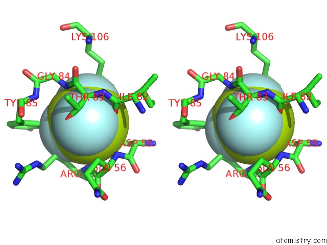
Fluorine binding site 3 out of 3 in 2vui
Go back to
Fluorine binding site 3 out
of 3 in the Crystal Structure of the Hupr Receiver Domain in Inhibitory Phospho-State
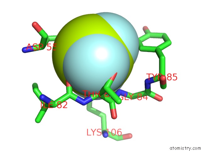
Mono view
Stereo pair view
Mono view
Stereo pair view
A full contact list of Fluorine with other atoms in the F binding
site number 3 of Crystal Structure of the Hupr Receiver Domain in Inhibitory Phospho-State within 5.0Å range:
|
Reference:
K.M.Davies,
E.D.Lowe,
C.Venien-Bryan,
L.N.Johnson.
The Hupr Receiver Domain Crystal Structure in Its Nonphospho and Inhibitory Phospho States. J.Mol.Biol. V. 385 51 2009.
ISSN: ISSN 0022-2836
PubMed: 18977359
DOI: 10.1016/J.JMB.2008.10.027
Page generated: Wed Jul 31 16:19:28 2024
ISSN: ISSN 0022-2836
PubMed: 18977359
DOI: 10.1016/J.JMB.2008.10.027
Last articles
Zn in 9J0NZn in 9J0O
Zn in 9J0P
Zn in 9FJX
Zn in 9EKB
Zn in 9C0F
Zn in 9CAH
Zn in 9CH0
Zn in 9CH3
Zn in 9CH1