Fluorine »
PDB 2weg-2x2f »
2wf8 »
Fluorine in PDB 2wf8: Structure of Beta-Phosphoglucomutase Inhibited with Glucose-6- Phosphate, Glucose-1-Phosphate and Beryllium Trifluoride
Enzymatic activity of Structure of Beta-Phosphoglucomutase Inhibited with Glucose-6- Phosphate, Glucose-1-Phosphate and Beryllium Trifluoride
All present enzymatic activity of Structure of Beta-Phosphoglucomutase Inhibited with Glucose-6- Phosphate, Glucose-1-Phosphate and Beryllium Trifluoride:
5.4.2.6;
5.4.2.6;
Protein crystallography data
The structure of Structure of Beta-Phosphoglucomutase Inhibited with Glucose-6- Phosphate, Glucose-1-Phosphate and Beryllium Trifluoride, PDB code: 2wf8
was solved by
M.W.Bowler,
N.J.Baxter,
C.E.Webster,
S.Pollard,
T.Alizadeh,
A.M.Hounslow,
M.J.Cliff,
W.Bermel,
N.H.Williams,
F.Hollfelder,
G.M.Blackburn,
J.P.Waltho,
with X-Ray Crystallography technique. A brief refinement statistics is given in the table below:
| Resolution Low / High (Å) | 20.00 / 1.20 |
| Space group | P 21 21 21 |
| Cell size a, b, c (Å), α, β, γ (°) | 37.300, 54.300, 104.200, 90.00, 90.00, 90.00 |
| R / Rfree (%) | 16.2 / 19.1 |
Other elements in 2wf8:
The structure of Structure of Beta-Phosphoglucomutase Inhibited with Glucose-6- Phosphate, Glucose-1-Phosphate and Beryllium Trifluoride also contains other interesting chemical elements:
| Magnesium | (Mg) | 1 atom |
| Sodium | (Na) | 2 atoms |
Fluorine Binding Sites:
The binding sites of Fluorine atom in the Structure of Beta-Phosphoglucomutase Inhibited with Glucose-6- Phosphate, Glucose-1-Phosphate and Beryllium Trifluoride
(pdb code 2wf8). This binding sites where shown within
5.0 Angstroms radius around Fluorine atom.
In total 3 binding sites of Fluorine where determined in the Structure of Beta-Phosphoglucomutase Inhibited with Glucose-6- Phosphate, Glucose-1-Phosphate and Beryllium Trifluoride, PDB code: 2wf8:
Jump to Fluorine binding site number: 1; 2; 3;
In total 3 binding sites of Fluorine where determined in the Structure of Beta-Phosphoglucomutase Inhibited with Glucose-6- Phosphate, Glucose-1-Phosphate and Beryllium Trifluoride, PDB code: 2wf8:
Jump to Fluorine binding site number: 1; 2; 3;
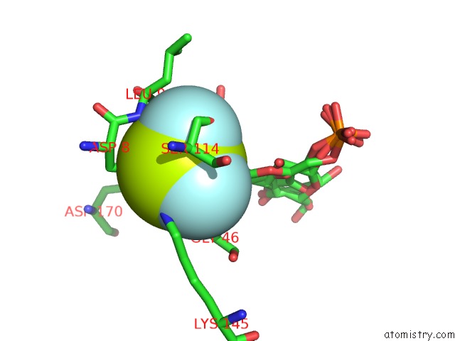
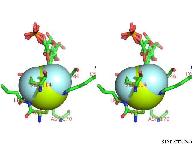
Fluorine binding site 1 out of 3 in 2wf8
Go back to
Fluorine binding site 1 out
of 3 in the Structure of Beta-Phosphoglucomutase Inhibited with Glucose-6- Phosphate, Glucose-1-Phosphate and Beryllium Trifluoride
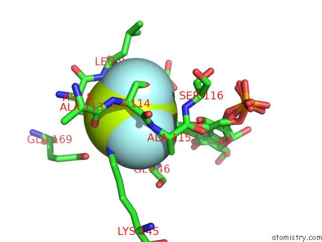
Mono view
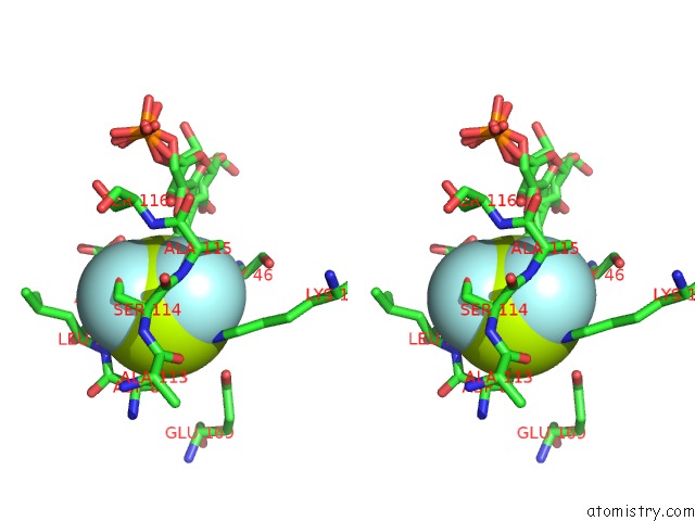
Stereo pair view
Mono view
Stereo pair view
A full contact list of Fluorine with other atoms in the F binding
site number 1 of Structure of Beta-Phosphoglucomutase Inhibited with Glucose-6- Phosphate, Glucose-1-Phosphate and Beryllium Trifluoride within 5.0Å range:
|
Fluorine binding site 2 out of 3 in 2wf8
Go back to
Fluorine binding site 2 out
of 3 in the Structure of Beta-Phosphoglucomutase Inhibited with Glucose-6- Phosphate, Glucose-1-Phosphate and Beryllium Trifluoride
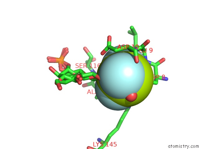
Mono view
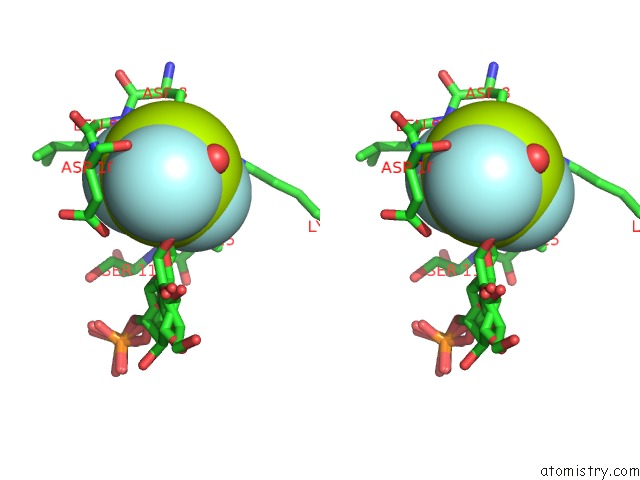
Stereo pair view
Mono view
Stereo pair view
A full contact list of Fluorine with other atoms in the F binding
site number 2 of Structure of Beta-Phosphoglucomutase Inhibited with Glucose-6- Phosphate, Glucose-1-Phosphate and Beryllium Trifluoride within 5.0Å range:
|
Fluorine binding site 3 out of 3 in 2wf8
Go back to
Fluorine binding site 3 out
of 3 in the Structure of Beta-Phosphoglucomutase Inhibited with Glucose-6- Phosphate, Glucose-1-Phosphate and Beryllium Trifluoride
Mono view
Stereo pair view
Mono view
Stereo pair view
A full contact list of Fluorine with other atoms in the F binding
site number 3 of Structure of Beta-Phosphoglucomutase Inhibited with Glucose-6- Phosphate, Glucose-1-Phosphate and Beryllium Trifluoride within 5.0Å range:
|
Reference:
J.L.Griffin,
M.W.Bowler,
N.J.Baxter,
K.N.Leigh,
H.R.Dannatt,
A.M.Hounslow,
G.M.Blackburn,
C.E.Webster,
M.J.Cliff,
J.P.Waltho.
Near Attack Conformers Dominate Beta-Phosphoglucomutase Complexes Where Geometry and Charge Distribution Reflect Those of Substrate. Proc. Natl. Acad. Sci. V. 109 6910 2012U.S.A..
ISSN: ESSN 1091-6490
PubMed: 22505741
DOI: 10.1073/PNAS.1116855109
Page generated: Wed Jul 31 16:25:07 2024
ISSN: ESSN 1091-6490
PubMed: 22505741
DOI: 10.1073/PNAS.1116855109
Last articles
Zn in 9MJ5Zn in 9HNW
Zn in 9G0L
Zn in 9FNE
Zn in 9DZN
Zn in 9E0I
Zn in 9D32
Zn in 9DAK
Zn in 8ZXC
Zn in 8ZUF