Fluorine »
PDB 2weg-2x2f »
2wxh »
Fluorine in PDB 2wxh: The Crystal Structure of the Murine Class Ia Pi 3-Kinase P110DELTA in Complex with SW14.
Enzymatic activity of The Crystal Structure of the Murine Class Ia Pi 3-Kinase P110DELTA in Complex with SW14.
All present enzymatic activity of The Crystal Structure of the Murine Class Ia Pi 3-Kinase P110DELTA in Complex with SW14.:
2.7.1.153;
2.7.1.153;
Protein crystallography data
The structure of The Crystal Structure of the Murine Class Ia Pi 3-Kinase P110DELTA in Complex with SW14., PDB code: 2wxh
was solved by
A.Berndt,
S.Miller,
O.Williams,
D.D.Lee,
B.T.Houseman,
J.I.Pacold,
F.Gorrec,
W.-C.Hon,
Y.Liu,
C.Rommel,
P.Gaillard,
T.Ruckle,
M.K.Schwarz,
K.M.Shokat,
J.P.Shaw,
R.L.Williams,
with X-Ray Crystallography technique. A brief refinement statistics is given in the table below:
| Resolution Low / High (Å) | 38.04 / 1.90 |
| Space group | C 1 2 1 |
| Cell size a, b, c (Å), α, β, γ (°) | 142.870, 65.010, 117.080, 90.00, 103.58, 90.00 |
| R / Rfree (%) | 21.7 / 24.5 |
Fluorine Binding Sites:
The binding sites of Fluorine atom in the The Crystal Structure of the Murine Class Ia Pi 3-Kinase P110DELTA in Complex with SW14.
(pdb code 2wxh). This binding sites where shown within
5.0 Angstroms radius around Fluorine atom.
In total only one binding site of Fluorine was determined in the The Crystal Structure of the Murine Class Ia Pi 3-Kinase P110DELTA in Complex with SW14., PDB code: 2wxh:
In total only one binding site of Fluorine was determined in the The Crystal Structure of the Murine Class Ia Pi 3-Kinase P110DELTA in Complex with SW14., PDB code: 2wxh:
Fluorine binding site 1 out of 1 in 2wxh
Go back to
Fluorine binding site 1 out
of 1 in the The Crystal Structure of the Murine Class Ia Pi 3-Kinase P110DELTA in Complex with SW14.
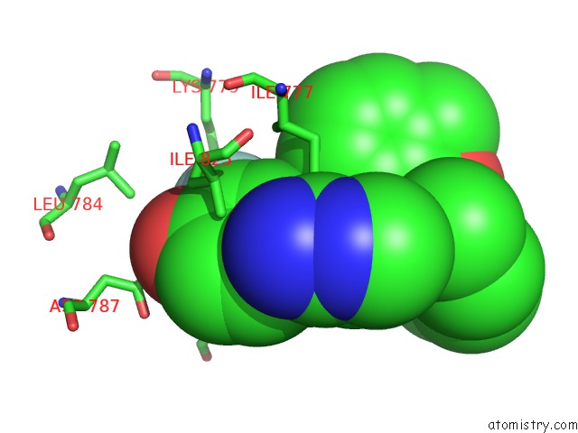
Mono view
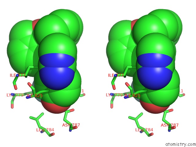
Stereo pair view
Mono view
Stereo pair view
A full contact list of Fluorine with other atoms in the F binding
site number 1 of The Crystal Structure of the Murine Class Ia Pi 3-Kinase P110DELTA in Complex with SW14. within 5.0Å range:
|
Reference:
A.Berndt,
S.Miller,
O.Williams,
D.D.Lee,
B.T.Houseman,
J.I.Pacold,
F.Gorrec,
W.-C.Hon,
Y.Liu,
C.Rommel,
P.Gaillard,
T.Ruckle,
M.K.Schwarz,
K.M.Shokat,
J.P.Shaw,
R.L.Williams.
The P110D Structure: Mechanisms For Selectivity and Potency of New Pi(3)K Inhibitors Nat.Chem.Biol. V. 6 117 2010.
ISSN: ISSN 1552-4450
PubMed: 20081827
DOI: 10.1038/NCHEMBIO.293
Page generated: Mon Jul 14 14:44:02 2025
ISSN: ISSN 1552-4450
PubMed: 20081827
DOI: 10.1038/NCHEMBIO.293
Last articles
F in 3WV1F in 3WGU
F in 3WRU
F in 3WPN
F in 3WK8
F in 3WIG
F in 3W9S
F in 3WFG
F in 3WFF
F in 3WF7