Fluorine »
PDB 2y1x-2z5z »
2yak »
Fluorine in PDB 2yak: Structure of Death-Associated Protein Kinase 1 (DAPK1) in Complex with A Ruthenium Octasporine Ligand (Osv)
Enzymatic activity of Structure of Death-Associated Protein Kinase 1 (DAPK1) in Complex with A Ruthenium Octasporine Ligand (Osv)
All present enzymatic activity of Structure of Death-Associated Protein Kinase 1 (DAPK1) in Complex with A Ruthenium Octasporine Ligand (Osv):
2.7.11.1;
2.7.11.1;
Protein crystallography data
The structure of Structure of Death-Associated Protein Kinase 1 (DAPK1) in Complex with A Ruthenium Octasporine Ligand (Osv), PDB code: 2yak
was solved by
L.Feng,
Y.Geisselbrecht,
S.Blanck,
A.Wilbuer,
G.E.Atilla-Gokcumen,
P.Filippakopoulos,
K.Kraeling,
M.A.Celik,
K.Harms,
J.Maksimoska,
R.Marmorstein,
G.Frenking,
S.Knapp,
L.-O.Essen,
E.Meggers,
with X-Ray Crystallography technique. A brief refinement statistics is given in the table below:
| Resolution Low / High (Å) | 24.53 / 2.20 |
| Space group | P 21 21 21 |
| Cell size a, b, c (Å), α, β, γ (°) | 50.300, 77.800, 111.300, 90.00, 90.00, 90.00 |
| R / Rfree (%) | 19.842 / 26.043 |
Other elements in 2yak:
The structure of Structure of Death-Associated Protein Kinase 1 (DAPK1) in Complex with A Ruthenium Octasporine Ligand (Osv) also contains other interesting chemical elements:
| Ruthenium | (Ru) | 1 atom |
Fluorine Binding Sites:
The binding sites of Fluorine atom in the Structure of Death-Associated Protein Kinase 1 (DAPK1) in Complex with A Ruthenium Octasporine Ligand (Osv)
(pdb code 2yak). This binding sites where shown within
5.0 Angstroms radius around Fluorine atom.
In total only one binding site of Fluorine was determined in the Structure of Death-Associated Protein Kinase 1 (DAPK1) in Complex with A Ruthenium Octasporine Ligand (Osv), PDB code: 2yak:
In total only one binding site of Fluorine was determined in the Structure of Death-Associated Protein Kinase 1 (DAPK1) in Complex with A Ruthenium Octasporine Ligand (Osv), PDB code: 2yak:
Fluorine binding site 1 out of 1 in 2yak
Go back to
Fluorine binding site 1 out
of 1 in the Structure of Death-Associated Protein Kinase 1 (DAPK1) in Complex with A Ruthenium Octasporine Ligand (Osv)
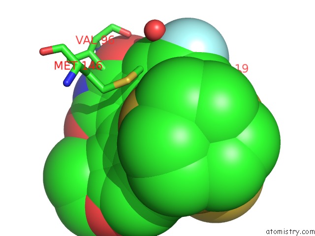
Mono view
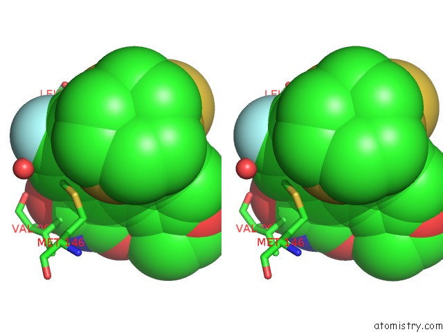
Stereo pair view
Mono view
Stereo pair view
A full contact list of Fluorine with other atoms in the F binding
site number 1 of Structure of Death-Associated Protein Kinase 1 (DAPK1) in Complex with A Ruthenium Octasporine Ligand (Osv) within 5.0Å range:
|
Reference:
L.Feng,
Y.Geisselbrecht,
S.Blanck,
A.Wilbuer,
G.E.Atilla-Gokcumen,
P.Filippakopoulos,
K.Kraeling,
M.A.Celik,
K.Harms,
J.Maksimoska,
R.Marmorstein,
G.Frenking,
S.Knapp,
L.O.Essen,
E.Meggers.
Structurally Sophisticated Octahedral Metal Complexes As Highly Selective Protein Kinase Inhibitors. J.Am.Chem.Soc. V. 133 5976 2011.
ISSN: ISSN 0002-7863
PubMed: 21446733
DOI: 10.1021/JA1112996
Page generated: Wed Jul 31 16:50:35 2024
ISSN: ISSN 0002-7863
PubMed: 21446733
DOI: 10.1021/JA1112996
Last articles
Zn in 9J0NZn in 9J0O
Zn in 9J0P
Zn in 9FJX
Zn in 9EKB
Zn in 9C0F
Zn in 9CAH
Zn in 9CH0
Zn in 9CH3
Zn in 9CH1