Fluorine »
PDB 2z78-3az8 »
3air »
Fluorine in PDB 3air: Crystal Structure of Beta-Glucosidase in Wheat Complexed with 2-Deoxy- 2-Fluoroglucoside and Dinitrophenol
Enzymatic activity of Crystal Structure of Beta-Glucosidase in Wheat Complexed with 2-Deoxy- 2-Fluoroglucoside and Dinitrophenol
All present enzymatic activity of Crystal Structure of Beta-Glucosidase in Wheat Complexed with 2-Deoxy- 2-Fluoroglucoside and Dinitrophenol:
3.2.1.21;
3.2.1.21;
Protein crystallography data
The structure of Crystal Structure of Beta-Glucosidase in Wheat Complexed with 2-Deoxy- 2-Fluoroglucoside and Dinitrophenol, PDB code: 3air
was solved by
M.Sue,
C.Nakamura,
T.Miyamoto,
S.Yajima,
with X-Ray Crystallography technique. A brief refinement statistics is given in the table below:
| Resolution Low / High (Å) | 39.69 / 2.00 |
| Space group | P 41 3 2 |
| Cell size a, b, c (Å), α, β, γ (°) | 194.448, 194.448, 194.448, 90.00, 90.00, 90.00 |
| R / Rfree (%) | 18 / 19.8 |
Fluorine Binding Sites:
The binding sites of Fluorine atom in the Crystal Structure of Beta-Glucosidase in Wheat Complexed with 2-Deoxy- 2-Fluoroglucoside and Dinitrophenol
(pdb code 3air). This binding sites where shown within
5.0 Angstroms radius around Fluorine atom.
In total only one binding site of Fluorine was determined in the Crystal Structure of Beta-Glucosidase in Wheat Complexed with 2-Deoxy- 2-Fluoroglucoside and Dinitrophenol, PDB code: 3air:
In total only one binding site of Fluorine was determined in the Crystal Structure of Beta-Glucosidase in Wheat Complexed with 2-Deoxy- 2-Fluoroglucoside and Dinitrophenol, PDB code: 3air:
Fluorine binding site 1 out of 1 in 3air
Go back to
Fluorine binding site 1 out
of 1 in the Crystal Structure of Beta-Glucosidase in Wheat Complexed with 2-Deoxy- 2-Fluoroglucoside and Dinitrophenol
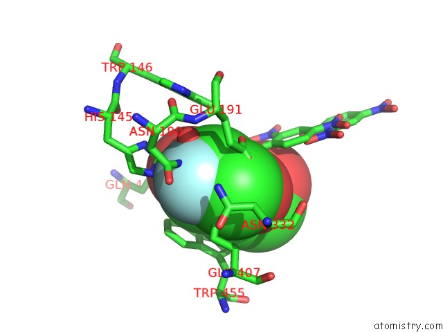
Mono view
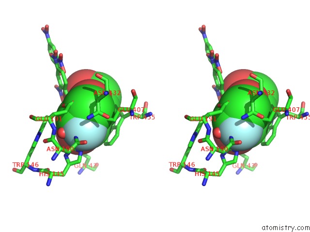
Stereo pair view
Mono view
Stereo pair view
A full contact list of Fluorine with other atoms in the F binding
site number 1 of Crystal Structure of Beta-Glucosidase in Wheat Complexed with 2-Deoxy- 2-Fluoroglucoside and Dinitrophenol within 5.0Å range:
|
Reference:
M.Sue,
C.Nakamura,
T.Miyamoto,
S.Yajima.
Active-Site Architecture of Benzoxazinone-Glucoside Beta-D-Glucosidases in Triticeae Plant Sci. V. 180 268 2011.
ISSN: ISSN 0168-9452
PubMed: 21421370
DOI: 10.1016/J.PLANTSCI.2010.09.001
Page generated: Wed Jul 31 17:08:20 2024
ISSN: ISSN 0168-9452
PubMed: 21421370
DOI: 10.1016/J.PLANTSCI.2010.09.001
Last articles
Zn in 9J0NZn in 9J0O
Zn in 9J0P
Zn in 9FJX
Zn in 9EKB
Zn in 9C0F
Zn in 9CAH
Zn in 9CH0
Zn in 9CH3
Zn in 9CH1