Fluorine in PDB 3cpc: Crystal Structure of the VEGFR2 Kinase Domain in Complex with A Pyridone Inhibitor
Enzymatic activity of Crystal Structure of the VEGFR2 Kinase Domain in Complex with A Pyridone Inhibitor
All present enzymatic activity of Crystal Structure of the VEGFR2 Kinase Domain in Complex with A Pyridone Inhibitor:
2.7.10.1;
Protein crystallography data
The structure of Crystal Structure of the VEGFR2 Kinase Domain in Complex with A Pyridone Inhibitor, PDB code: 3cpc
was solved by
D.A.Whittington,
A.M.Long,
P.Rose,
Y.Gu,
H.Zhao,
with X-Ray Crystallography technique. A brief refinement statistics is given in the table below:
Resolution Low / High (Å)
|
30.00 /
2.40
|
Space group
|
P 1 21 1
|
Cell size a, b, c (Å), α, β, γ (°)
|
55.361,
66.865,
89.858,
90.00,
93.35,
90.00
|
R / Rfree (%)
|
21.8 /
27.2
|
Fluorine Binding Sites:
The binding sites of Fluorine atom in the Crystal Structure of the VEGFR2 Kinase Domain in Complex with A Pyridone Inhibitor
(pdb code 3cpc). This binding sites where shown within
5.0 Angstroms radius around Fluorine atom.
In total 6 binding sites of Fluorine where determined in the
Crystal Structure of the VEGFR2 Kinase Domain in Complex with A Pyridone Inhibitor, PDB code: 3cpc:
Jump to Fluorine binding site number:
1;
2;
3;
4;
5;
6;
Fluorine binding site 1 out
of 6 in 3cpc
Go back to
Fluorine Binding Sites List in 3cpc
Fluorine binding site 1 out
of 6 in the Crystal Structure of the VEGFR2 Kinase Domain in Complex with A Pyridone Inhibitor
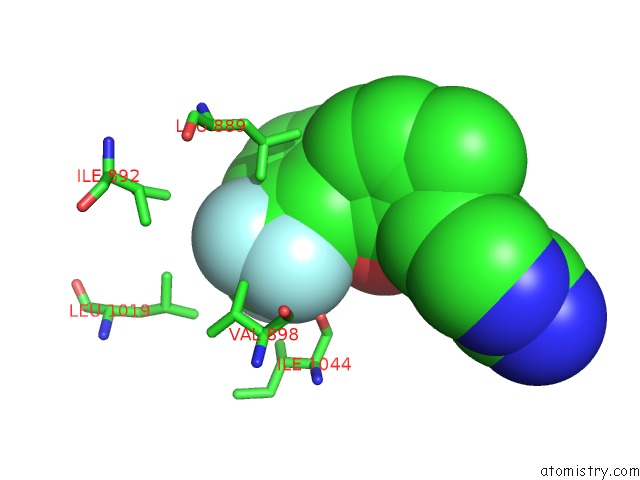
Mono view
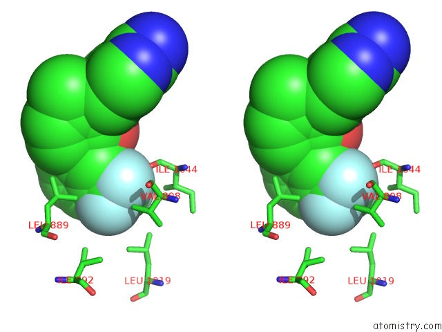
Stereo pair view
|
A full contact list of Fluorine with other atoms in the F binding
site number 1 of Crystal Structure of the VEGFR2 Kinase Domain in Complex with A Pyridone Inhibitor within 5.0Å range:
probe
|
atom
|
residue
|
distance (Å)
|
B
|
Occ
|
A:F1
b:28.2
occ:1.00
|
F2
|
A:C521
|
0.0
|
28.2
|
1.0
|
C21
|
A:C521
|
1.3
|
27.0
|
1.0
|
F1
|
A:C521
|
2.1
|
26.9
|
1.0
|
F3
|
A:C521
|
2.2
|
26.9
|
1.0
|
C17
|
A:C521
|
2.4
|
26.8
|
1.0
|
C18
|
A:C521
|
3.0
|
26.5
|
1.0
|
CD2
|
A:LEU889
|
3.5
|
34.6
|
1.0
|
C16
|
A:C521
|
3.5
|
26.1
|
1.0
|
CG1
|
A:VAL898
|
3.7
|
27.0
|
1.0
|
C19
|
A:C521
|
4.3
|
26.3
|
1.0
|
CD2
|
A:LEU1019
|
4.5
|
27.1
|
1.0
|
CD1
|
A:ILE892
|
4.6
|
35.6
|
1.0
|
C15
|
A:C521
|
4.6
|
25.8
|
1.0
|
CG
|
A:LEU889
|
4.7
|
34.3
|
1.0
|
CD1
|
A:LEU1019
|
4.8
|
27.3
|
1.0
|
O
|
A:ILE1044
|
4.9
|
27.6
|
1.0
|
C20
|
A:C521
|
5.0
|
26.4
|
1.0
|
|
Fluorine binding site 2 out
of 6 in 3cpc
Go back to
Fluorine Binding Sites List in 3cpc
Fluorine binding site 2 out
of 6 in the Crystal Structure of the VEGFR2 Kinase Domain in Complex with A Pyridone Inhibitor
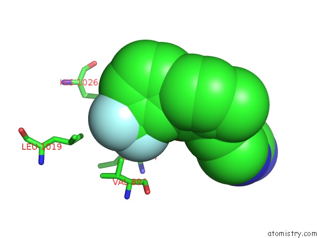
Mono view
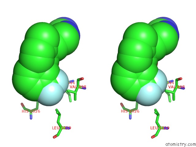
Stereo pair view
|
A full contact list of Fluorine with other atoms in the F binding
site number 2 of Crystal Structure of the VEGFR2 Kinase Domain in Complex with A Pyridone Inhibitor within 5.0Å range:
probe
|
atom
|
residue
|
distance (Å)
|
B
|
Occ
|
A:F1
b:26.9
occ:1.00
|
F3
|
A:C521
|
0.0
|
26.9
|
1.0
|
C21
|
A:C521
|
1.3
|
27.0
|
1.0
|
F2
|
A:C521
|
2.2
|
28.2
|
1.0
|
F1
|
A:C521
|
2.2
|
26.9
|
1.0
|
C17
|
A:C521
|
2.3
|
26.8
|
1.0
|
C18
|
A:C521
|
2.9
|
26.5
|
1.0
|
C16
|
A:C521
|
3.3
|
26.1
|
1.0
|
CD2
|
A:HIS1026
|
3.7
|
34.5
|
1.0
|
NE2
|
A:HIS1026
|
3.8
|
34.1
|
1.0
|
C
|
A:CYS1045
|
3.9
|
31.5
|
1.0
|
CG2
|
A:ILE1044
|
3.9
|
27.1
|
1.0
|
O
|
A:CYS1045
|
3.9
|
31.7
|
1.0
|
O
|
A:ILE1044
|
4.1
|
27.6
|
1.0
|
CB
|
A:ASP1046
|
4.2
|
34.7
|
1.0
|
CA
|
A:CYS1045
|
4.2
|
30.3
|
1.0
|
C19
|
A:C521
|
4.2
|
26.3
|
1.0
|
N
|
A:ASP1046
|
4.3
|
32.8
|
1.0
|
C
|
A:ILE1044
|
4.4
|
28.0
|
1.0
|
N
|
A:CYS1045
|
4.5
|
28.8
|
1.0
|
C15
|
A:C521
|
4.5
|
25.8
|
1.0
|
CD1
|
A:LEU1019
|
4.8
|
27.3
|
1.0
|
CG1
|
A:VAL898
|
4.8
|
27.0
|
1.0
|
CA
|
A:ASP1046
|
4.8
|
34.4
|
1.0
|
CB
|
A:ILE1044
|
4.8
|
27.1
|
1.0
|
C20
|
A:C521
|
4.9
|
26.4
|
1.0
|
CG
|
A:HIS1026
|
4.9
|
34.6
|
1.0
|
OD2
|
A:ASP1046
|
5.0
|
37.4
|
1.0
|
|
Fluorine binding site 3 out
of 6 in 3cpc
Go back to
Fluorine Binding Sites List in 3cpc
Fluorine binding site 3 out
of 6 in the Crystal Structure of the VEGFR2 Kinase Domain in Complex with A Pyridone Inhibitor
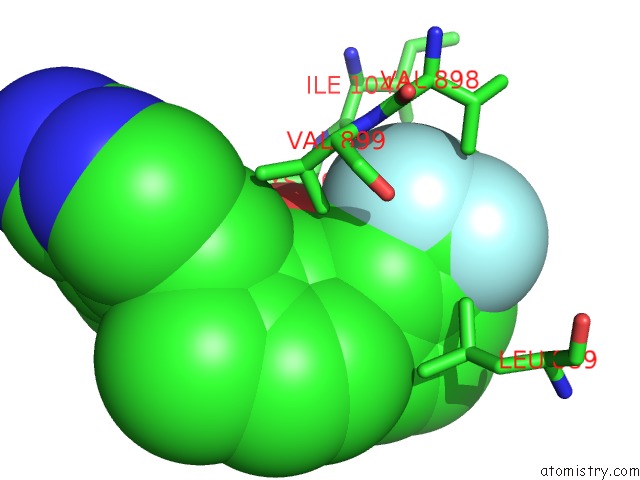
Mono view
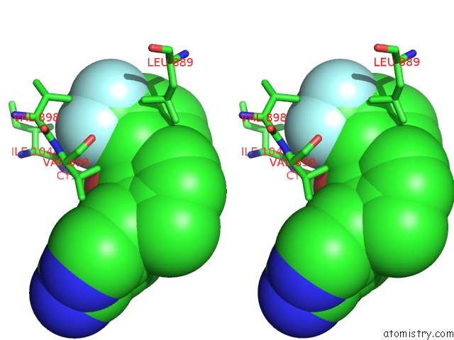
Stereo pair view
|
A full contact list of Fluorine with other atoms in the F binding
site number 3 of Crystal Structure of the VEGFR2 Kinase Domain in Complex with A Pyridone Inhibitor within 5.0Å range:
probe
|
atom
|
residue
|
distance (Å)
|
B
|
Occ
|
A:F1
b:26.9
occ:1.00
|
F1
|
A:C521
|
0.0
|
26.9
|
1.0
|
C21
|
A:C521
|
1.3
|
27.0
|
1.0
|
F2
|
A:C521
|
2.1
|
28.2
|
1.0
|
F3
|
A:C521
|
2.2
|
26.9
|
1.0
|
C17
|
A:C521
|
2.4
|
26.8
|
1.0
|
C16
|
A:C521
|
2.7
|
26.1
|
1.0
|
O
|
A:ILE1044
|
3.0
|
27.6
|
1.0
|
CG1
|
A:VAL898
|
3.4
|
27.0
|
1.0
|
CG1
|
A:VAL899
|
3.5
|
26.3
|
1.0
|
C18
|
A:C521
|
3.7
|
26.5
|
1.0
|
C
|
A:ILE1044
|
3.8
|
28.0
|
1.0
|
CA
|
A:CYS1045
|
3.9
|
30.3
|
1.0
|
N
|
A:VAL899
|
4.1
|
27.0
|
1.0
|
C15
|
A:C521
|
4.1
|
25.8
|
1.0
|
N
|
A:CYS1045
|
4.2
|
28.8
|
1.0
|
CD2
|
A:LEU889
|
4.3
|
34.6
|
1.0
|
C
|
A:CYS1045
|
4.4
|
31.5
|
1.0
|
O
|
A:VAL899
|
4.5
|
27.1
|
1.0
|
CB
|
A:VAL899
|
4.5
|
26.8
|
1.0
|
CG2
|
A:ILE1044
|
4.6
|
27.1
|
1.0
|
O1
|
A:C521
|
4.6
|
25.1
|
1.0
|
CB
|
A:VAL898
|
4.6
|
26.9
|
1.0
|
N
|
A:ASP1046
|
4.8
|
32.8
|
1.0
|
CA
|
A:VAL899
|
4.8
|
26.7
|
1.0
|
C
|
A:VAL898
|
4.8
|
27.0
|
1.0
|
C19
|
A:C521
|
4.8
|
26.3
|
1.0
|
CA
|
A:VAL898
|
4.8
|
26.9
|
1.0
|
CB
|
A:ILE1044
|
4.8
|
27.1
|
1.0
|
O
|
A:CYS1045
|
4.8
|
31.7
|
1.0
|
CA
|
A:ILE1044
|
4.9
|
27.3
|
1.0
|
N4
|
A:C521
|
5.0
|
26.0
|
1.0
|
C20
|
A:C521
|
5.0
|
26.4
|
1.0
|
|
Fluorine binding site 4 out
of 6 in 3cpc
Go back to
Fluorine Binding Sites List in 3cpc
Fluorine binding site 4 out
of 6 in the Crystal Structure of the VEGFR2 Kinase Domain in Complex with A Pyridone Inhibitor
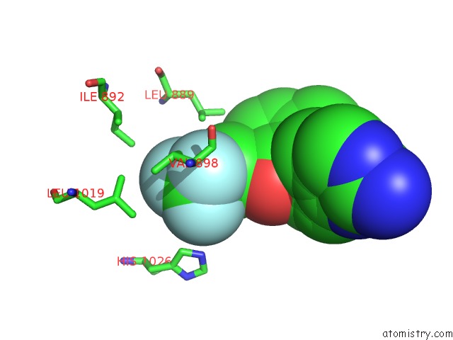
Mono view
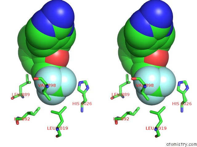
Stereo pair view
|
A full contact list of Fluorine with other atoms in the F binding
site number 4 of Crystal Structure of the VEGFR2 Kinase Domain in Complex with A Pyridone Inhibitor within 5.0Å range:
probe
|
atom
|
residue
|
distance (Å)
|
B
|
Occ
|
B:F2
b:30.9
occ:1.00
|
F2
|
B:C522
|
0.0
|
30.9
|
1.0
|
C21
|
B:C522
|
1.3
|
29.9
|
1.0
|
F1
|
B:C522
|
2.2
|
31.4
|
1.0
|
F3
|
B:C522
|
2.2
|
30.3
|
1.0
|
C17
|
B:C522
|
2.3
|
29.4
|
1.0
|
C18
|
B:C522
|
2.8
|
29.1
|
1.0
|
C16
|
B:C522
|
3.6
|
29.1
|
1.0
|
C19
|
B:C522
|
4.1
|
28.9
|
1.0
|
CG1
|
B:VAL898
|
4.2
|
37.2
|
1.0
|
CD1
|
B:ILE892
|
4.3
|
45.8
|
1.0
|
CD2
|
B:LEU889
|
4.4
|
45.5
|
1.0
|
CD2
|
B:LEU1019
|
4.5
|
38.9
|
1.0
|
CD1
|
B:LEU1019
|
4.7
|
38.9
|
1.0
|
C15
|
B:C522
|
4.7
|
28.3
|
1.0
|
C20
|
B:C522
|
4.9
|
28.9
|
1.0
|
CD2
|
B:HIS1026
|
4.9
|
39.9
|
1.0
|
|
Fluorine binding site 5 out
of 6 in 3cpc
Go back to
Fluorine Binding Sites List in 3cpc
Fluorine binding site 5 out
of 6 in the Crystal Structure of the VEGFR2 Kinase Domain in Complex with A Pyridone Inhibitor
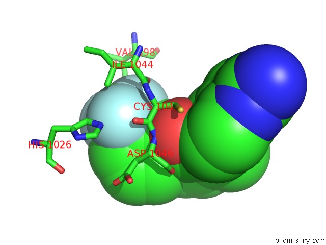
Mono view
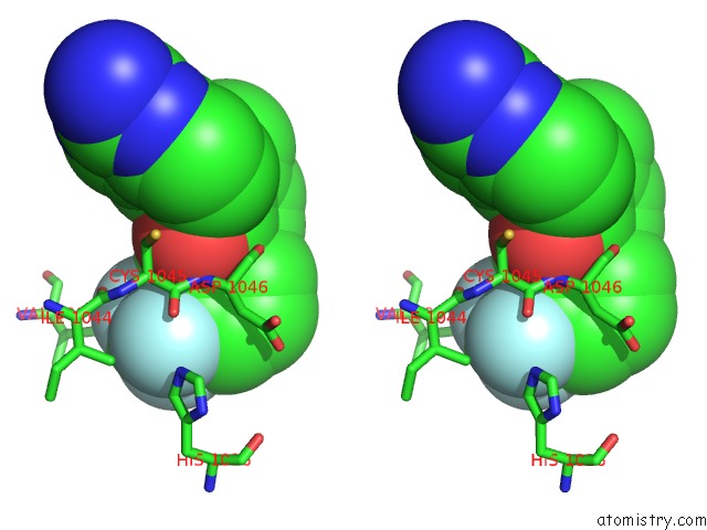
Stereo pair view
|
A full contact list of Fluorine with other atoms in the F binding
site number 5 of Crystal Structure of the VEGFR2 Kinase Domain in Complex with A Pyridone Inhibitor within 5.0Å range:
probe
|
atom
|
residue
|
distance (Å)
|
B
|
Occ
|
B:F2
b:30.3
occ:1.00
|
F3
|
B:C522
|
0.0
|
30.3
|
1.0
|
C21
|
B:C522
|
1.3
|
29.9
|
1.0
|
F1
|
B:C522
|
2.2
|
31.4
|
1.0
|
F2
|
B:C522
|
2.2
|
30.9
|
1.0
|
C17
|
B:C522
|
2.3
|
29.4
|
1.0
|
C18
|
B:C522
|
3.1
|
29.1
|
1.0
|
C16
|
B:C522
|
3.2
|
29.1
|
1.0
|
C
|
B:CYS1045
|
3.5
|
34.7
|
1.0
|
CG2
|
B:ILE1044
|
3.5
|
31.8
|
1.0
|
O
|
B:CYS1045
|
3.6
|
34.5
|
1.0
|
NE2
|
B:HIS1026
|
3.6
|
39.2
|
1.0
|
CD2
|
B:HIS1026
|
3.6
|
39.9
|
1.0
|
CA
|
B:CYS1045
|
3.7
|
34.3
|
1.0
|
O
|
B:ILE1044
|
3.8
|
32.7
|
1.0
|
N
|
B:ASP1046
|
3.9
|
35.8
|
1.0
|
N
|
B:CYS1045
|
4.0
|
33.5
|
1.0
|
C
|
B:ILE1044
|
4.0
|
33.1
|
1.0
|
CB
|
B:ASP1046
|
4.1
|
37.1
|
1.0
|
C19
|
B:C522
|
4.3
|
28.9
|
1.0
|
C15
|
B:C522
|
4.4
|
28.3
|
1.0
|
CB
|
B:ILE1044
|
4.6
|
32.9
|
1.0
|
CA
|
B:ASP1046
|
4.6
|
36.9
|
1.0
|
CE1
|
B:HIS1026
|
4.8
|
39.4
|
1.0
|
CG1
|
B:VAL898
|
4.9
|
37.2
|
1.0
|
C20
|
B:C522
|
4.9
|
28.9
|
1.0
|
CG
|
B:HIS1026
|
4.9
|
40.1
|
1.0
|
CA
|
B:ILE1044
|
4.9
|
32.9
|
1.0
|
|
Fluorine binding site 6 out
of 6 in 3cpc
Go back to
Fluorine Binding Sites List in 3cpc
Fluorine binding site 6 out
of 6 in the Crystal Structure of the VEGFR2 Kinase Domain in Complex with A Pyridone Inhibitor
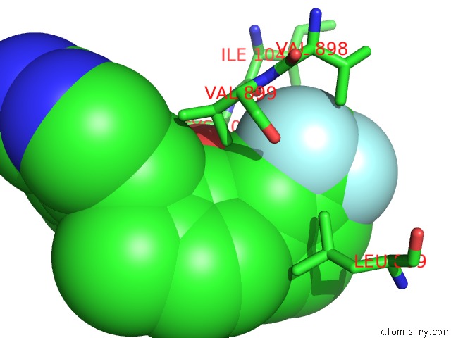
Mono view
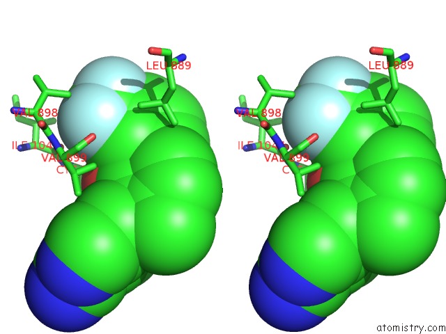
Stereo pair view
|
A full contact list of Fluorine with other atoms in the F binding
site number 6 of Crystal Structure of the VEGFR2 Kinase Domain in Complex with A Pyridone Inhibitor within 5.0Å range:
probe
|
atom
|
residue
|
distance (Å)
|
B
|
Occ
|
B:F2
b:31.4
occ:1.00
|
F1
|
B:C522
|
0.0
|
31.4
|
1.0
|
C21
|
B:C522
|
1.3
|
29.9
|
1.0
|
F3
|
B:C522
|
2.2
|
30.3
|
1.0
|
F2
|
B:C522
|
2.2
|
30.9
|
1.0
|
C17
|
B:C522
|
2.3
|
29.4
|
1.0
|
C16
|
B:C522
|
2.7
|
29.1
|
1.0
|
CG1
|
B:VAL898
|
3.3
|
37.2
|
1.0
|
O
|
B:ILE1044
|
3.3
|
32.7
|
1.0
|
C18
|
B:C522
|
3.6
|
29.1
|
1.0
|
CG1
|
B:VAL899
|
3.7
|
37.7
|
1.0
|
C
|
B:ILE1044
|
4.1
|
33.1
|
1.0
|
C15
|
B:C522
|
4.1
|
28.3
|
1.0
|
CD2
|
B:LEU889
|
4.1
|
45.5
|
1.0
|
CA
|
B:CYS1045
|
4.2
|
34.3
|
1.0
|
N
|
B:VAL899
|
4.3
|
37.7
|
1.0
|
O
|
B:VAL899
|
4.3
|
38.8
|
1.0
|
N
|
B:CYS1045
|
4.5
|
33.5
|
1.0
|
CG2
|
B:ILE1044
|
4.6
|
31.8
|
1.0
|
C
|
B:CYS1045
|
4.6
|
34.7
|
1.0
|
CB
|
B:VAL898
|
4.6
|
36.9
|
1.0
|
C19
|
B:C522
|
4.7
|
28.9
|
1.0
|
CB
|
B:VAL899
|
4.8
|
38.0
|
1.0
|
O1
|
B:C522
|
4.8
|
27.1
|
1.0
|
C
|
B:VAL898
|
4.9
|
37.1
|
1.0
|
CA
|
B:VAL899
|
4.9
|
38.1
|
1.0
|
CA
|
B:VAL898
|
4.9
|
37.0
|
1.0
|
C20
|
B:C522
|
4.9
|
28.9
|
1.0
|
N
|
B:ASP1046
|
4.9
|
35.8
|
1.0
|
|
Reference:
E.Hu,
A.Tasker,
R.D.White,
R.K.Kunz,
J.Human,
N.Chen,
R.Burli,
R.Hungate,
P.Novak,
A.Itano,
X.Zhang,
V.Yu,
Y.Nguyen,
Y.Tudor,
M.Plant,
S.Flynn,
Y.Xu,
K.L.Meagher,
D.A.Whittington,
G.Y.Ng.
Discovery of Aryl Aminoquinazoline Pyridones As Potent, Selective, and Orally Efficacious Inhibitors of Receptor Tyrosine Kinase C-Kit. J.Med.Chem. V. 51 3065 2008.
ISSN: ISSN 0022-2623
PubMed: 18447379
DOI: 10.1021/JM800188G
Page generated: Wed Jul 31 17:33:20 2024
|