Fluorine »
PDB 3ccw-3d39 »
3cpo »
Fluorine in PDB 3cpo: Crystal Structure of Ketosteroid Isomerase D40N with Bound 2- Fluorophenol
Enzymatic activity of Crystal Structure of Ketosteroid Isomerase D40N with Bound 2- Fluorophenol
All present enzymatic activity of Crystal Structure of Ketosteroid Isomerase D40N with Bound 2- Fluorophenol:
5.3.3.1;
5.3.3.1;
Protein crystallography data
The structure of Crystal Structure of Ketosteroid Isomerase D40N with Bound 2- Fluorophenol, PDB code: 3cpo
was solved by
J.M.M.Caaveiro,
B.Pybus,
D.Ringe,
G.Petsko,
with X-Ray Crystallography technique. A brief refinement statistics is given in the table below:
| Resolution Low / High (Å) | 36.19 / 1.24 |
| Space group | C 2 2 21 |
| Cell size a, b, c (Å), α, β, γ (°) | 35.347, 95.013, 72.376, 90.00, 90.00, 90.00 |
| R / Rfree (%) | 16.6 / 20 |
Fluorine Binding Sites:
The binding sites of Fluorine atom in the Crystal Structure of Ketosteroid Isomerase D40N with Bound 2- Fluorophenol
(pdb code 3cpo). This binding sites where shown within
5.0 Angstroms radius around Fluorine atom.
In total only one binding site of Fluorine was determined in the Crystal Structure of Ketosteroid Isomerase D40N with Bound 2- Fluorophenol, PDB code: 3cpo:
In total only one binding site of Fluorine was determined in the Crystal Structure of Ketosteroid Isomerase D40N with Bound 2- Fluorophenol, PDB code: 3cpo:
Fluorine binding site 1 out of 1 in 3cpo
Go back to
Fluorine binding site 1 out
of 1 in the Crystal Structure of Ketosteroid Isomerase D40N with Bound 2- Fluorophenol
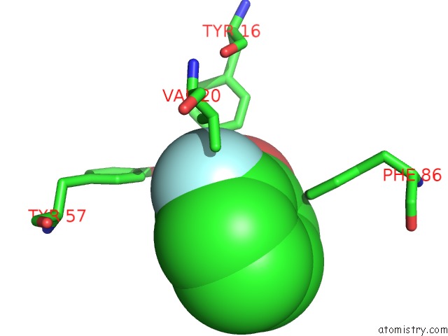
Mono view
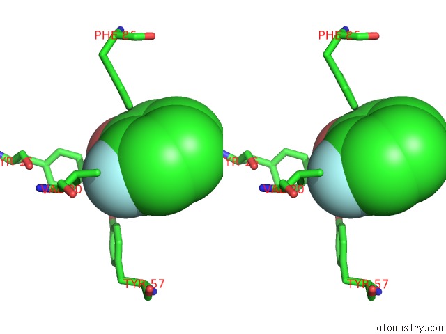
Stereo pair view
Mono view
Stereo pair view
A full contact list of Fluorine with other atoms in the F binding
site number 1 of Crystal Structure of Ketosteroid Isomerase D40N with Bound 2- Fluorophenol within 5.0Å range:
|
Reference:
P.A.Sigala,
D.A.Kraut,
J.M.M.Caaveiro,
B.Pybus,
E.A.Ruben,
D.Ringe,
G.A.Petsko,
D.Herschlag.
Testing Geometrical Discrimination Within An Enzyme Active Site: Constrained Hydrogen Bonding in the Ketosteroid Isomerase Oxyanion Hole J.Am.Chem.Soc. V. 130 13696 2008.
ISSN: ISSN 0002-7863
PubMed: 18808119
DOI: 10.1021/JA803928M
Page generated: Mon Jul 14 15:40:15 2025
ISSN: ISSN 0002-7863
PubMed: 18808119
DOI: 10.1021/JA803928M
Last articles
Fe in 2YXOFe in 2YRS
Fe in 2YXC
Fe in 2YNM
Fe in 2YVJ
Fe in 2YP1
Fe in 2YU2
Fe in 2YU1
Fe in 2YQB
Fe in 2YOO