Fluorine »
PDB 3ccw-3d39 »
3cpw »
Fluorine in PDB 3cpw: The Structure of the Antibiotic Linezolid Bound to the Large Ribosomal Subunit of Haloarcula Marismortui
Protein crystallography data
The structure of The Structure of the Antibiotic Linezolid Bound to the Large Ribosomal Subunit of Haloarcula Marismortui, PDB code: 3cpw
was solved by
J.A.Ippolito,
Z.K.Kanyo,
D.Wang,
F.J.Franceschi,
P.B.Moore,
T.A.Steitz,
E.M.Duffy,
with X-Ray Crystallography technique. A brief refinement statistics is given in the table below:
| Resolution Low / High (Å) | 40.00 / 2.70 |
| Space group | C 2 2 21 |
| Cell size a, b, c (Å), α, β, γ (°) | 211.730, 298.580, 575.290, 90.00, 90.00, 90.00 |
| R / Rfree (%) | n/a / n/a |
Other elements in 3cpw:
The structure of The Structure of the Antibiotic Linezolid Bound to the Large Ribosomal Subunit of Haloarcula Marismortui also contains other interesting chemical elements:
| Strontium | (Sr) | 108 atoms |
| Magnesium | (Mg) | 92 atoms |
| Potassium | (K) | 2 atoms |
| Cadmium | (Cd) | 5 atoms |
| Chlorine | (Cl) | 22 atoms |
| Sodium | (Na) | 75 atoms |
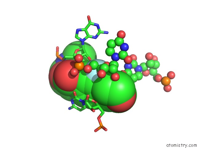
Fluorine Binding Sites:
The binding sites of Fluorine atom in the The Structure of the Antibiotic Linezolid Bound to the Large Ribosomal Subunit of Haloarcula Marismortui
(pdb code 3cpw). This binding sites where shown within
5.0 Angstroms radius around Fluorine atom.
In total only one binding site of Fluorine was determined in the The Structure of the Antibiotic Linezolid Bound to the Large Ribosomal Subunit of Haloarcula Marismortui, PDB code: 3cpw:
In total only one binding site of Fluorine was determined in the The Structure of the Antibiotic Linezolid Bound to the Large Ribosomal Subunit of Haloarcula Marismortui, PDB code: 3cpw:
Fluorine binding site 1 out of 1 in 3cpw
Go back to
Fluorine binding site 1 out
of 1 in the The Structure of the Antibiotic Linezolid Bound to the Large Ribosomal Subunit of Haloarcula Marismortui
Mono view
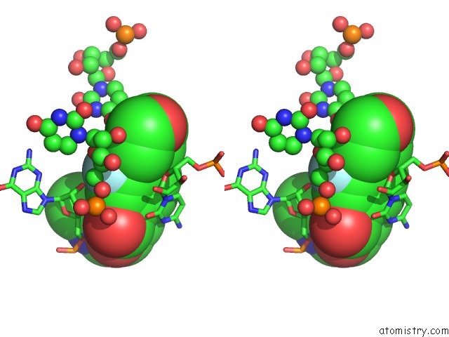
Stereo pair view
Mono view
Stereo pair view
A full contact list of Fluorine with other atoms in the F binding
site number 1 of The Structure of the Antibiotic Linezolid Bound to the Large Ribosomal Subunit of Haloarcula Marismortui within 5.0Å range:
|
Reference:
J.A.Ippolito,
Z.K.Kanyo,
D.Wang,
F.J.Franceschi,
P.B.Moore,
T.A.Steitz,
E.M.Duffy.
Crystal Structure of the Oxazolidinone Antibiotic Linezolid Bound to the 50S Ribosomal Subunit J.Med.Chem. V. 51 3353 2008.
ISSN: ISSN 0022-2623
PubMed: 18494460
DOI: 10.1021/JM800379D
Page generated: Wed Jul 31 17:35:26 2024
ISSN: ISSN 0022-2623
PubMed: 18494460
DOI: 10.1021/JM800379D
Last articles
Zn in 9JYWZn in 9IR4
Zn in 9IR3
Zn in 9GMX
Zn in 9GMW
Zn in 9JEJ
Zn in 9ERF
Zn in 9ERE
Zn in 9EGV
Zn in 9EGW