Fluorine »
PDB 3d3e-3du8 »
3d6u »
Fluorine in PDB 3d6u: Crystal Structure of 4-(Trifluoromethyldiazirinyl) Phenylalanyl-Trna Synthetase
Enzymatic activity of Crystal Structure of 4-(Trifluoromethyldiazirinyl) Phenylalanyl-Trna Synthetase
All present enzymatic activity of Crystal Structure of 4-(Trifluoromethyldiazirinyl) Phenylalanyl-Trna Synthetase:
6.1.1.1;
6.1.1.1;
Protein crystallography data
The structure of Crystal Structure of 4-(Trifluoromethyldiazirinyl) Phenylalanyl-Trna Synthetase, PDB code: 3d6u
was solved by
W.Liu,
E.Tippmann,
A.V.Mack,
P.G.Schultz,
with X-Ray Crystallography technique. A brief refinement statistics is given in the table below:
| Resolution Low / High (Å) | 45.98 / 2.20 |
| Space group | P 43 21 2 |
| Cell size a, b, c (Å), α, β, γ (°) | 102.779, 102.779, 70.780, 90.00, 90.00, 90.00 |
| R / Rfree (%) | 20.1 / 27.4 |
Fluorine Binding Sites:
The binding sites of Fluorine atom in the Crystal Structure of 4-(Trifluoromethyldiazirinyl) Phenylalanyl-Trna Synthetase
(pdb code 3d6u). This binding sites where shown within
5.0 Angstroms radius around Fluorine atom.
In total 3 binding sites of Fluorine where determined in the Crystal Structure of 4-(Trifluoromethyldiazirinyl) Phenylalanyl-Trna Synthetase, PDB code: 3d6u:
Jump to Fluorine binding site number: 1; 2; 3;
In total 3 binding sites of Fluorine where determined in the Crystal Structure of 4-(Trifluoromethyldiazirinyl) Phenylalanyl-Trna Synthetase, PDB code: 3d6u:
Jump to Fluorine binding site number: 1; 2; 3;
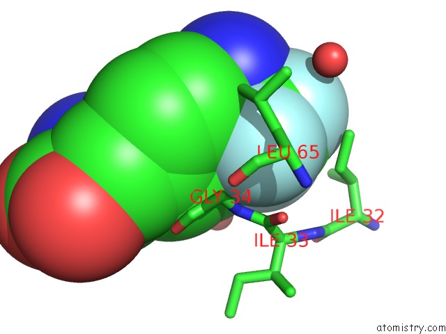
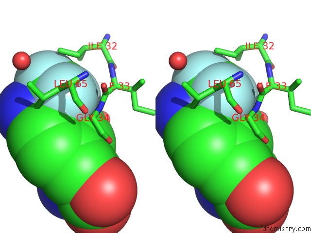
Fluorine binding site 1 out of 3 in 3d6u
Go back to
Fluorine binding site 1 out
of 3 in the Crystal Structure of 4-(Trifluoromethyldiazirinyl) Phenylalanyl-Trna Synthetase
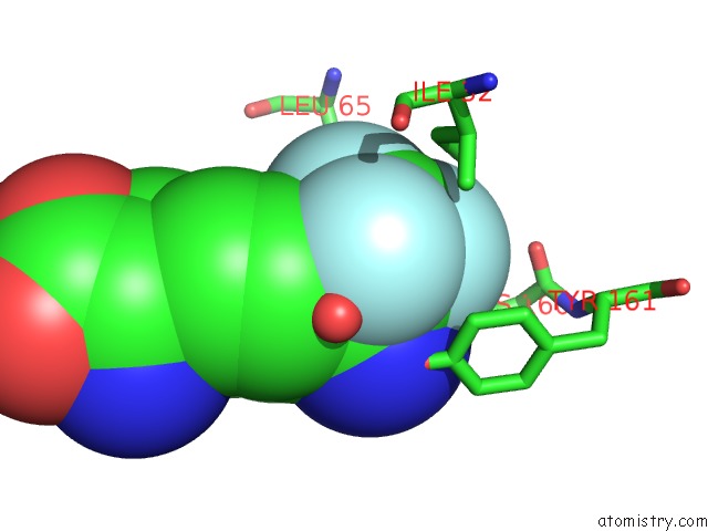
Mono view
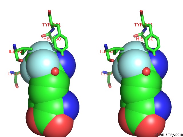
Stereo pair view
Mono view
Stereo pair view
A full contact list of Fluorine with other atoms in the F binding
site number 1 of Crystal Structure of 4-(Trifluoromethyldiazirinyl) Phenylalanyl-Trna Synthetase within 5.0Å range:
|
Fluorine binding site 2 out of 3 in 3d6u
Go back to
Fluorine binding site 2 out
of 3 in the Crystal Structure of 4-(Trifluoromethyldiazirinyl) Phenylalanyl-Trna Synthetase
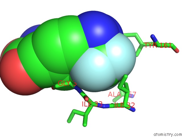
Mono view
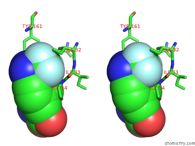
Stereo pair view
Mono view
Stereo pair view
A full contact list of Fluorine with other atoms in the F binding
site number 2 of Crystal Structure of 4-(Trifluoromethyldiazirinyl) Phenylalanyl-Trna Synthetase within 5.0Å range:
|
Fluorine binding site 3 out of 3 in 3d6u
Go back to
Fluorine binding site 3 out
of 3 in the Crystal Structure of 4-(Trifluoromethyldiazirinyl) Phenylalanyl-Trna Synthetase
Mono view
Stereo pair view
Mono view
Stereo pair view
A full contact list of Fluorine with other atoms in the F binding
site number 3 of Crystal Structure of 4-(Trifluoromethyldiazirinyl) Phenylalanyl-Trna Synthetase within 5.0Å range:
|
Reference:
E.Tippmann,
W.Liu,
D.Summerer,
A.V.Mack,
P.G.Schultz.
A Genetically Encoded Diazirine Photocrosslinker in Escherichia Coli Chembiochem V. 8 2210 2007.
ISSN: ISSN 1439-4227
PubMed: 18000916
DOI: 10.1002/CBIC.200700460
Page generated: Wed Jul 31 17:51:31 2024
ISSN: ISSN 1439-4227
PubMed: 18000916
DOI: 10.1002/CBIC.200700460
Last articles
Zn in 9J0NZn in 9J0O
Zn in 9J0P
Zn in 9FJX
Zn in 9EKB
Zn in 9C0F
Zn in 9CAH
Zn in 9CH0
Zn in 9CH3
Zn in 9CH1