Fluorine »
PDB 3d3e-3du8 »
3d7m »
Fluorine in PDB 3d7m: Crystal Structure of the G Protein Fast-Exchange Double Mutant I56C/Q333C
Protein crystallography data
The structure of Crystal Structure of the G Protein Fast-Exchange Double Mutant I56C/Q333C, PDB code: 3d7m
was solved by
M.A.Funk,
A.M.Preininger,
W.M.Oldham,
S.M.Meier,
H.E.Hamm,
T.M.Iverson,
with X-Ray Crystallography technique. A brief refinement statistics is given in the table below:
| Resolution Low / High (Å) | 50.00 / 2.90 |
| Space group | P 43 21 2 |
| Cell size a, b, c (Å), α, β, γ (°) | 79.659, 79.659, 114.587, 90.00, 90.00, 90.00 |
| R / Rfree (%) | 24.9 / 29.4 |
Other elements in 3d7m:
The structure of Crystal Structure of the G Protein Fast-Exchange Double Mutant I56C/Q333C also contains other interesting chemical elements:
| Magnesium | (Mg) | 1 atom |
| Aluminium | (Al) | 1 atom |
Fluorine Binding Sites:
The binding sites of Fluorine atom in the Crystal Structure of the G Protein Fast-Exchange Double Mutant I56C/Q333C
(pdb code 3d7m). This binding sites where shown within
5.0 Angstroms radius around Fluorine atom.
In total 4 binding sites of Fluorine where determined in the Crystal Structure of the G Protein Fast-Exchange Double Mutant I56C/Q333C, PDB code: 3d7m:
Jump to Fluorine binding site number: 1; 2; 3; 4;
In total 4 binding sites of Fluorine where determined in the Crystal Structure of the G Protein Fast-Exchange Double Mutant I56C/Q333C, PDB code: 3d7m:
Jump to Fluorine binding site number: 1; 2; 3; 4;
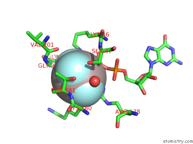
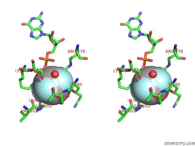
Fluorine binding site 1 out of 4 in 3d7m
Go back to
Fluorine binding site 1 out
of 4 in the Crystal Structure of the G Protein Fast-Exchange Double Mutant I56C/Q333C
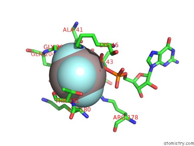
Mono view
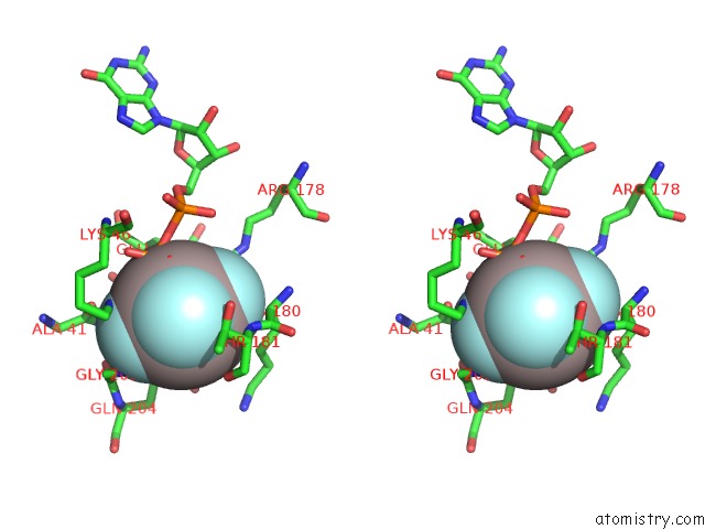
Stereo pair view
Mono view
Stereo pair view
A full contact list of Fluorine with other atoms in the F binding
site number 1 of Crystal Structure of the G Protein Fast-Exchange Double Mutant I56C/Q333C within 5.0Å range:
|
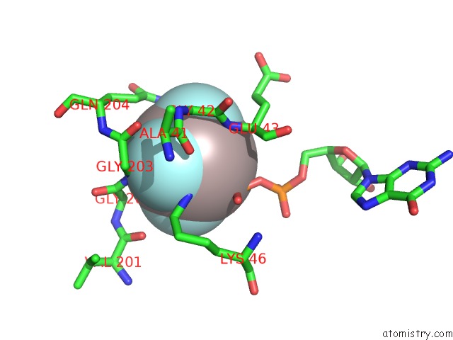
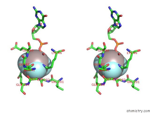
Fluorine binding site 2 out of 4 in 3d7m
Go back to
Fluorine binding site 2 out
of 4 in the Crystal Structure of the G Protein Fast-Exchange Double Mutant I56C/Q333C
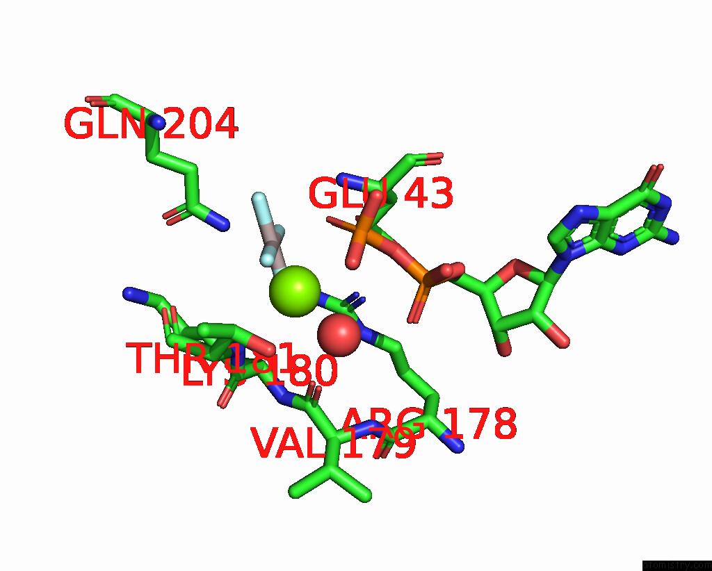
Mono view
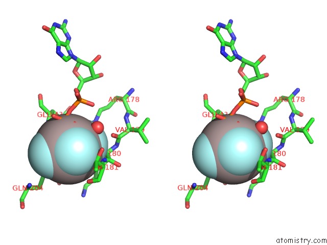
Stereo pair view
Mono view
Stereo pair view
A full contact list of Fluorine with other atoms in the F binding
site number 2 of Crystal Structure of the G Protein Fast-Exchange Double Mutant I56C/Q333C within 5.0Å range:
|
Fluorine binding site 3 out of 4 in 3d7m
Go back to
Fluorine binding site 3 out
of 4 in the Crystal Structure of the G Protein Fast-Exchange Double Mutant I56C/Q333C
Mono view
Stereo pair view
Mono view
Stereo pair view
A full contact list of Fluorine with other atoms in the F binding
site number 3 of Crystal Structure of the G Protein Fast-Exchange Double Mutant I56C/Q333C within 5.0Å range:
|
Fluorine binding site 4 out of 4 in 3d7m
Go back to
Fluorine binding site 4 out
of 4 in the Crystal Structure of the G Protein Fast-Exchange Double Mutant I56C/Q333C
Mono view
Stereo pair view
Mono view
Stereo pair view
A full contact list of Fluorine with other atoms in the F binding
site number 4 of Crystal Structure of the G Protein Fast-Exchange Double Mutant I56C/Q333C within 5.0Å range:
|
Reference:
A.M.Preininger,
M.A.Funk,
W.M.Oldham,
S.M.Meier,
C.A.Johnston,
S.Adhikary,
A.J.Kimple,
D.P.Siderovski,
H.E.Hamm,
T.M.Iverson.
Helix Dipole Movement and Conformational Variability Contribute to Allosteric Gdp Release in Galphai Subunits. Biochemistry V. 48 2630 2009.
ISSN: ISSN 0006-2960
PubMed: 19222191
DOI: 10.1021/BI801853A
Page generated: Mon Jul 14 15:48:32 2025
ISSN: ISSN 0006-2960
PubMed: 19222191
DOI: 10.1021/BI801853A
Last articles
Fe in 2YXOFe in 2YRS
Fe in 2YXC
Fe in 2YNM
Fe in 2YVJ
Fe in 2YP1
Fe in 2YU2
Fe in 2YU1
Fe in 2YQB
Fe in 2YOO