Fluorine »
PDB 3d3e-3du8 »
3dd2 »
Fluorine in PDB 3dd2: Crystal Structure of An Rna Aptamer Bound to Human Thrombin
Enzymatic activity of Crystal Structure of An Rna Aptamer Bound to Human Thrombin
All present enzymatic activity of Crystal Structure of An Rna Aptamer Bound to Human Thrombin:
3.4.21.5;
3.4.21.5;
Protein crystallography data
The structure of Crystal Structure of An Rna Aptamer Bound to Human Thrombin, PDB code: 3dd2
was solved by
S.B.Long,
B.A.Sullenger,
with X-Ray Crystallography technique. A brief refinement statistics is given in the table below:
| Resolution Low / High (Å) | 30.35 / 1.90 |
| Space group | P 21 21 2 |
| Cell size a, b, c (Å), α, β, γ (°) | 83.766, 134.017, 44.033, 90.00, 90.00, 90.00 |
| R / Rfree (%) | 20.4 / 25.4 |
Other elements in 3dd2:
The structure of Crystal Structure of An Rna Aptamer Bound to Human Thrombin also contains other interesting chemical elements:
| Magnesium | (Mg) | 2 atoms |
Fluorine Binding Sites:
The binding sites of Fluorine atom in the Crystal Structure of An Rna Aptamer Bound to Human Thrombin
(pdb code 3dd2). This binding sites where shown within
5.0 Angstroms radius around Fluorine atom.
In total 10 binding sites of Fluorine where determined in the Crystal Structure of An Rna Aptamer Bound to Human Thrombin, PDB code: 3dd2:
Jump to Fluorine binding site number: 1; 2; 3; 4; 5; 6; 7; 8; 9; 10;
In total 10 binding sites of Fluorine where determined in the Crystal Structure of An Rna Aptamer Bound to Human Thrombin, PDB code: 3dd2:
Jump to Fluorine binding site number: 1; 2; 3; 4; 5; 6; 7; 8; 9; 10;
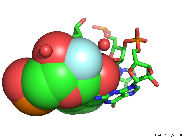
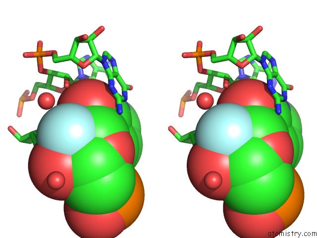
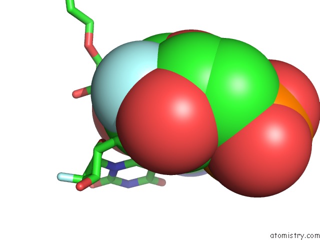
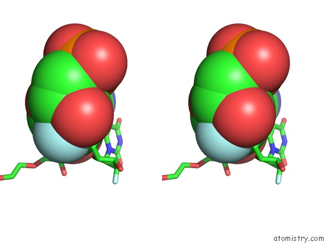
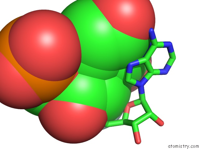
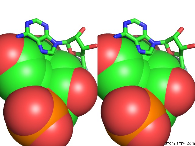
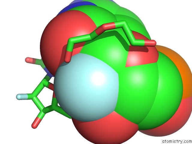
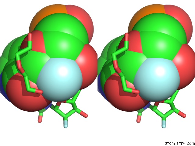
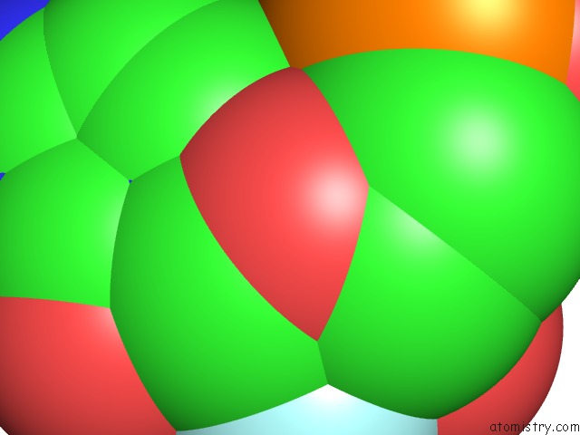
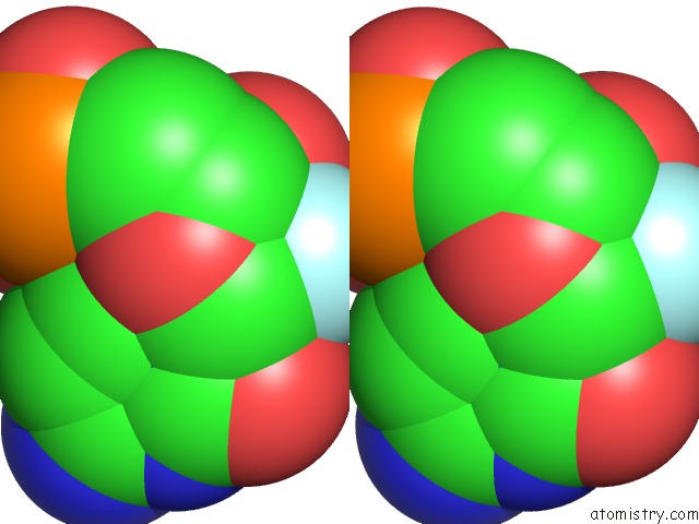
Fluorine binding site 1 out of 10 in 3dd2
Go back to
Fluorine binding site 1 out
of 10 in the Crystal Structure of An Rna Aptamer Bound to Human Thrombin
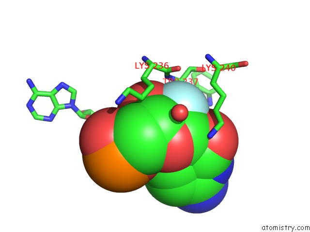
Mono view
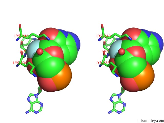
Stereo pair view
Mono view
Stereo pair view
A full contact list of Fluorine with other atoms in the F binding
site number 1 of Crystal Structure of An Rna Aptamer Bound to Human Thrombin within 5.0Å range:
|
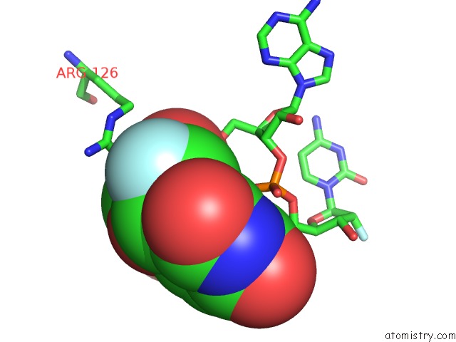
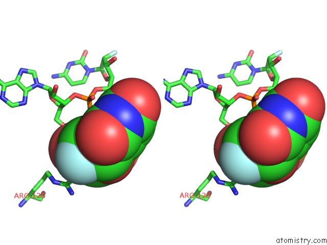
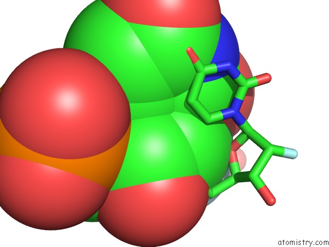
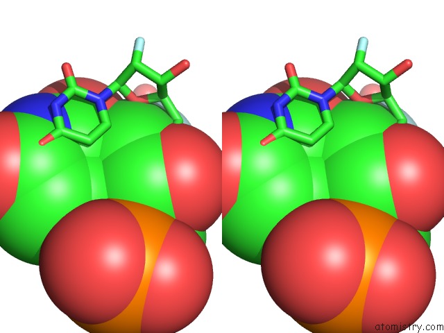
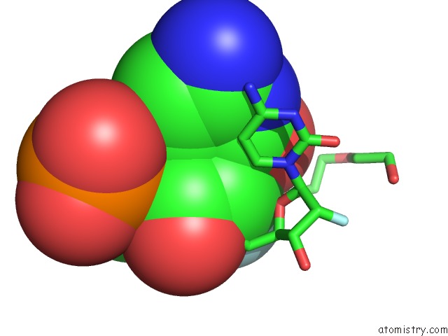
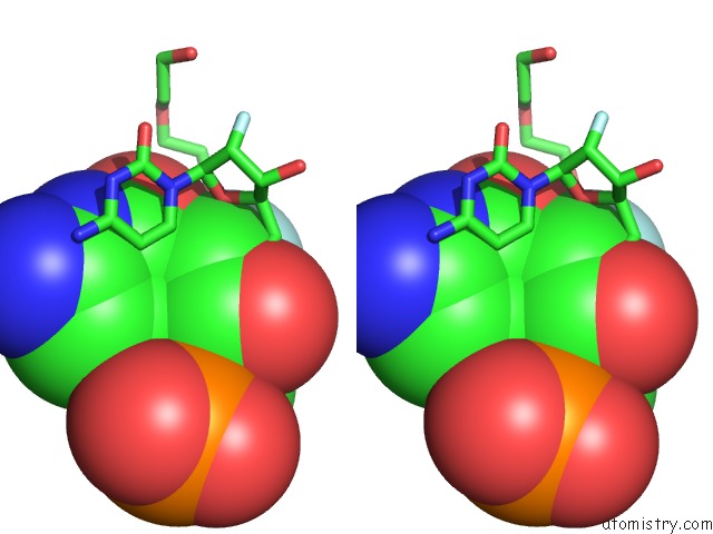
Fluorine binding site 2 out of 10 in 3dd2
Go back to
Fluorine binding site 2 out
of 10 in the Crystal Structure of An Rna Aptamer Bound to Human Thrombin
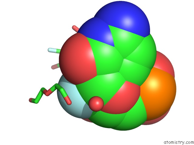
Mono view
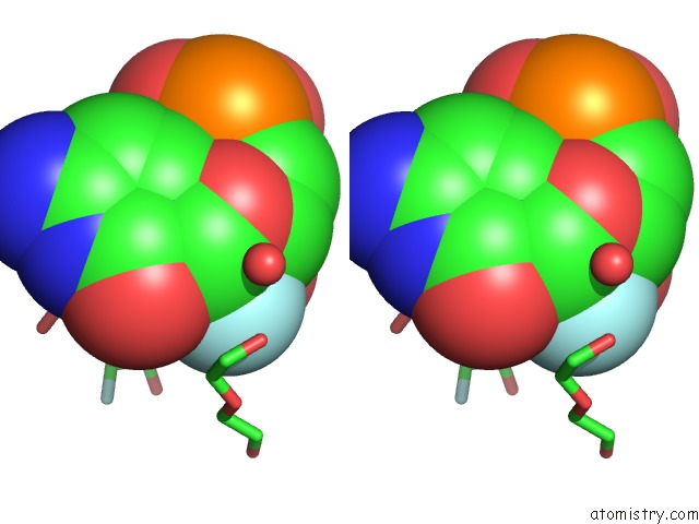
Stereo pair view
Mono view
Stereo pair view
A full contact list of Fluorine with other atoms in the F binding
site number 2 of Crystal Structure of An Rna Aptamer Bound to Human Thrombin within 5.0Å range:
|
Fluorine binding site 3 out of 10 in 3dd2
Go back to
Fluorine binding site 3 out
of 10 in the Crystal Structure of An Rna Aptamer Bound to Human Thrombin
Mono view
Stereo pair view
Mono view
Stereo pair view
A full contact list of Fluorine with other atoms in the F binding
site number 3 of Crystal Structure of An Rna Aptamer Bound to Human Thrombin within 5.0Å range:
|
Fluorine binding site 4 out of 10 in 3dd2
Go back to
Fluorine binding site 4 out
of 10 in the Crystal Structure of An Rna Aptamer Bound to Human Thrombin
Mono view
Stereo pair view
Mono view
Stereo pair view
A full contact list of Fluorine with other atoms in the F binding
site number 4 of Crystal Structure of An Rna Aptamer Bound to Human Thrombin within 5.0Å range:
|
Fluorine binding site 5 out of 10 in 3dd2
Go back to
Fluorine binding site 5 out
of 10 in the Crystal Structure of An Rna Aptamer Bound to Human Thrombin
Mono view
Stereo pair view
Mono view
Stereo pair view
A full contact list of Fluorine with other atoms in the F binding
site number 5 of Crystal Structure of An Rna Aptamer Bound to Human Thrombin within 5.0Å range:
|
Fluorine binding site 6 out of 10 in 3dd2
Go back to
Fluorine binding site 6 out
of 10 in the Crystal Structure of An Rna Aptamer Bound to Human Thrombin
Mono view
Stereo pair view
Mono view
Stereo pair view
A full contact list of Fluorine with other atoms in the F binding
site number 6 of Crystal Structure of An Rna Aptamer Bound to Human Thrombin within 5.0Å range:
|
Fluorine binding site 7 out of 10 in 3dd2
Go back to
Fluorine binding site 7 out
of 10 in the Crystal Structure of An Rna Aptamer Bound to Human Thrombin
Mono view
Stereo pair view
Mono view
Stereo pair view
A full contact list of Fluorine with other atoms in the F binding
site number 7 of Crystal Structure of An Rna Aptamer Bound to Human Thrombin within 5.0Å range:
|
Fluorine binding site 8 out of 10 in 3dd2
Go back to
Fluorine binding site 8 out
of 10 in the Crystal Structure of An Rna Aptamer Bound to Human Thrombin
Mono view
Stereo pair view
Mono view
Stereo pair view
A full contact list of Fluorine with other atoms in the F binding
site number 8 of Crystal Structure of An Rna Aptamer Bound to Human Thrombin within 5.0Å range:
|
Fluorine binding site 9 out of 10 in 3dd2
Go back to
Fluorine binding site 9 out
of 10 in the Crystal Structure of An Rna Aptamer Bound to Human Thrombin
Mono view
Stereo pair view
Mono view
Stereo pair view
A full contact list of Fluorine with other atoms in the F binding
site number 9 of Crystal Structure of An Rna Aptamer Bound to Human Thrombin within 5.0Å range:
|
Fluorine binding site 10 out of 10 in 3dd2
Go back to
Fluorine binding site 10 out
of 10 in the Crystal Structure of An Rna Aptamer Bound to Human Thrombin
Mono view
Stereo pair view
Mono view
Stereo pair view
A full contact list of Fluorine with other atoms in the F binding
site number 10 of Crystal Structure of An Rna Aptamer Bound to Human Thrombin within 5.0Å range:
|
Reference:
S.B.Long,
M.B.Long,
R.R.White,
B.A.Sullenger.
Crystal Structure of An Rna Aptamer Bound to Thrombin. Rna V. 14 2504 2008.
ISSN: ISSN 1355-8382
PubMed: 18971322
DOI: 10.1261/RNA.1239308
Page generated: Wed Jul 31 17:53:59 2024
ISSN: ISSN 1355-8382
PubMed: 18971322
DOI: 10.1261/RNA.1239308
Last articles
Zn in 9J0NZn in 9J0O
Zn in 9J0P
Zn in 9FJX
Zn in 9EKB
Zn in 9C0F
Zn in 9CAH
Zn in 9CH0
Zn in 9CH3
Zn in 9CH1