Fluorine »
PDB 3d3e-3du8 »
3dm2 »
Fluorine in PDB 3dm2: Crystal Structure of Hiv-1 K103N Mutant Reverse Transcriptase in Complex with GW564511.
Enzymatic activity of Crystal Structure of Hiv-1 K103N Mutant Reverse Transcriptase in Complex with GW564511.
All present enzymatic activity of Crystal Structure of Hiv-1 K103N Mutant Reverse Transcriptase in Complex with GW564511.:
2.7.7.49; 2.7.7.7; 3.1.26.4;
2.7.7.49; 2.7.7.7; 3.1.26.4;
Protein crystallography data
The structure of Crystal Structure of Hiv-1 K103N Mutant Reverse Transcriptase in Complex with GW564511., PDB code: 3dm2
was solved by
J.Ren,
P.P.Chamberlain,
D.K.Stammers,
with X-Ray Crystallography technique. A brief refinement statistics is given in the table below:
| Resolution Low / High (Å) | 29.54 / 3.10 |
| Space group | P 21 21 21 |
| Cell size a, b, c (Å), α, β, γ (°) | 135.910, 109.210, 71.410, 90.00, 90.00, 90.00 |
| R / Rfree (%) | 22.2 / 30.7 |
Other elements in 3dm2:
The structure of Crystal Structure of Hiv-1 K103N Mutant Reverse Transcriptase in Complex with GW564511. also contains other interesting chemical elements:
| Chlorine | (Cl) | 1 atom |
Fluorine Binding Sites:
The binding sites of Fluorine atom in the Crystal Structure of Hiv-1 K103N Mutant Reverse Transcriptase in Complex with GW564511.
(pdb code 3dm2). This binding sites where shown within
5.0 Angstroms radius around Fluorine atom.
In total 4 binding sites of Fluorine where determined in the Crystal Structure of Hiv-1 K103N Mutant Reverse Transcriptase in Complex with GW564511., PDB code: 3dm2:
Jump to Fluorine binding site number: 1; 2; 3; 4;
In total 4 binding sites of Fluorine where determined in the Crystal Structure of Hiv-1 K103N Mutant Reverse Transcriptase in Complex with GW564511., PDB code: 3dm2:
Jump to Fluorine binding site number: 1; 2; 3; 4;
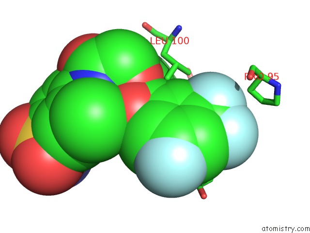
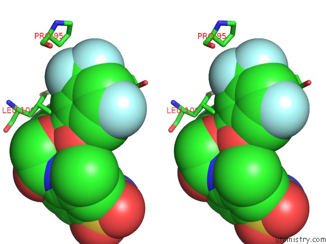
Fluorine binding site 1 out of 4 in 3dm2
Go back to
Fluorine binding site 1 out
of 4 in the Crystal Structure of Hiv-1 K103N Mutant Reverse Transcriptase in Complex with GW564511.
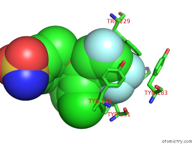
Mono view
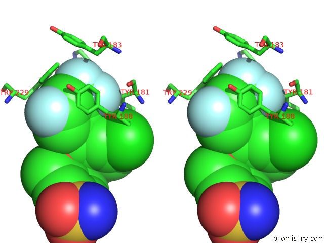
Stereo pair view
Mono view
Stereo pair view
A full contact list of Fluorine with other atoms in the F binding
site number 1 of Crystal Structure of Hiv-1 K103N Mutant Reverse Transcriptase in Complex with GW564511. within 5.0Å range:
|
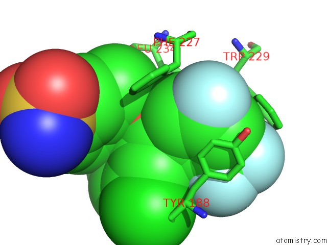
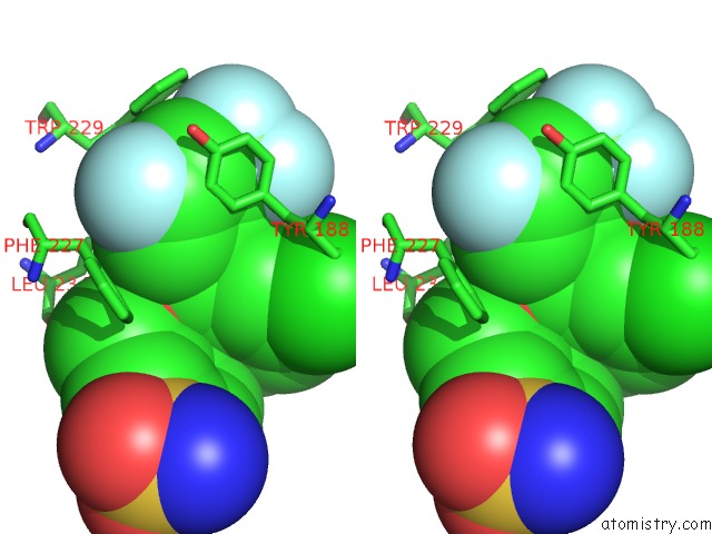
Fluorine binding site 2 out of 4 in 3dm2
Go back to
Fluorine binding site 2 out
of 4 in the Crystal Structure of Hiv-1 K103N Mutant Reverse Transcriptase in Complex with GW564511.
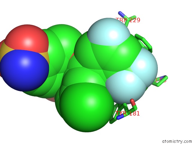
Mono view
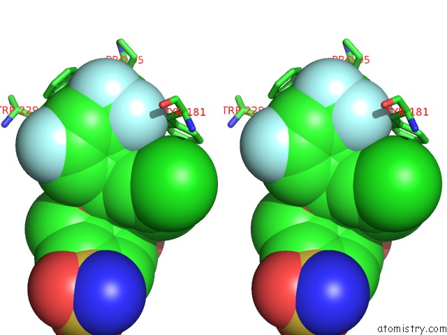
Stereo pair view
Mono view
Stereo pair view
A full contact list of Fluorine with other atoms in the F binding
site number 2 of Crystal Structure of Hiv-1 K103N Mutant Reverse Transcriptase in Complex with GW564511. within 5.0Å range:
|
Fluorine binding site 3 out of 4 in 3dm2
Go back to
Fluorine binding site 3 out
of 4 in the Crystal Structure of Hiv-1 K103N Mutant Reverse Transcriptase in Complex with GW564511.
Mono view
Stereo pair view
Mono view
Stereo pair view
A full contact list of Fluorine with other atoms in the F binding
site number 3 of Crystal Structure of Hiv-1 K103N Mutant Reverse Transcriptase in Complex with GW564511. within 5.0Å range:
|
Fluorine binding site 4 out of 4 in 3dm2
Go back to
Fluorine binding site 4 out
of 4 in the Crystal Structure of Hiv-1 K103N Mutant Reverse Transcriptase in Complex with GW564511.
Mono view
Stereo pair view
Mono view
Stereo pair view
A full contact list of Fluorine with other atoms in the F binding
site number 4 of Crystal Structure of Hiv-1 K103N Mutant Reverse Transcriptase in Complex with GW564511. within 5.0Å range:
|
Reference:
J.Ren,
P.P.Chamberlain,
A.Stamp,
S.A.Short,
K.L.Weaver,
K.R.Romines,
R.Hazen,
A.Freeman,
R.G.Ferris,
C.W.Andrews,
L.Boone,
J.H.Chan,
D.K.Stammers.
Structural Basis For the Improved Drug Resistance Profile of New Generation Benzophenone Non-Nucleoside Hiv-1 Reverse Transcriptase Inhibitors. J.Med.Chem. V. 51 5000 2008.
ISSN: ISSN 0022-2623
PubMed: 18665583
DOI: 10.1021/JM8004493
Page generated: Wed Jul 31 18:00:16 2024
ISSN: ISSN 0022-2623
PubMed: 18665583
DOI: 10.1021/JM8004493
Last articles
Zn in 9J0NZn in 9J0O
Zn in 9J0P
Zn in 9FJX
Zn in 9EKB
Zn in 9C0F
Zn in 9CAH
Zn in 9CH0
Zn in 9CH3
Zn in 9CH1