Fluorine »
PDB 3dv3-3ev4 »
3el7 »
Fluorine in PDB 3el7: Crystal Structure of C-Src in Complex with Pyrazolopyrimidine 3
Enzymatic activity of Crystal Structure of C-Src in Complex with Pyrazolopyrimidine 3
All present enzymatic activity of Crystal Structure of C-Src in Complex with Pyrazolopyrimidine 3:
2.7.10.2;
2.7.10.2;
Protein crystallography data
The structure of Crystal Structure of C-Src in Complex with Pyrazolopyrimidine 3, PDB code: 3el7
was solved by
A.C.Dar,
M.S.Lopez,
K.M.Shokat,
with X-Ray Crystallography technique. A brief refinement statistics is given in the table below:
| Resolution Low / High (Å) | 30.00 / 2.80 |
| Space group | P 1 21 1 |
| Cell size a, b, c (Å), α, β, γ (°) | 42.360, 63.130, 56.050, 90.00, 91.90, 90.00 |
| R / Rfree (%) | 22 / 28.8 |
Fluorine Binding Sites:
The binding sites of Fluorine atom in the Crystal Structure of C-Src in Complex with Pyrazolopyrimidine 3
(pdb code 3el7). This binding sites where shown within
5.0 Angstroms radius around Fluorine atom.
In total 3 binding sites of Fluorine where determined in the Crystal Structure of C-Src in Complex with Pyrazolopyrimidine 3, PDB code: 3el7:
Jump to Fluorine binding site number: 1; 2; 3;
In total 3 binding sites of Fluorine where determined in the Crystal Structure of C-Src in Complex with Pyrazolopyrimidine 3, PDB code: 3el7:
Jump to Fluorine binding site number: 1; 2; 3;
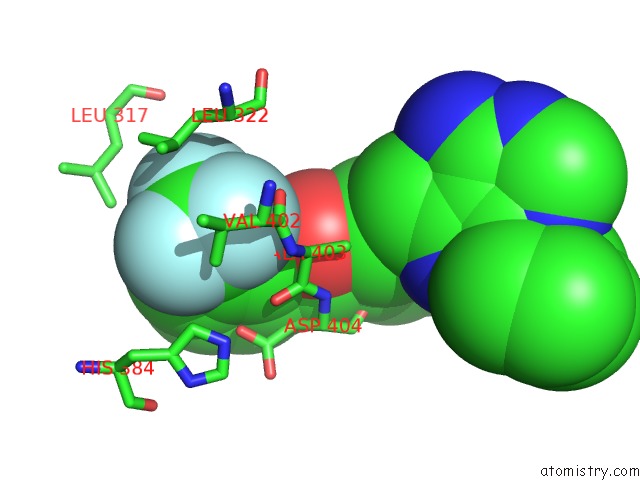
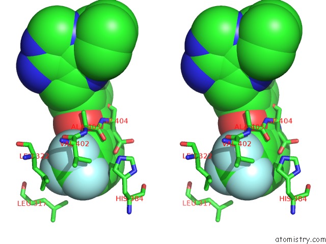
Fluorine binding site 1 out of 3 in 3el7
Go back to
Fluorine binding site 1 out
of 3 in the Crystal Structure of C-Src in Complex with Pyrazolopyrimidine 3
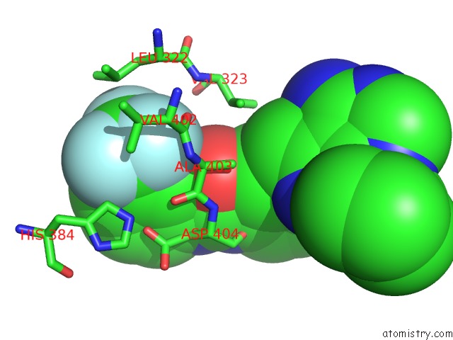
Mono view
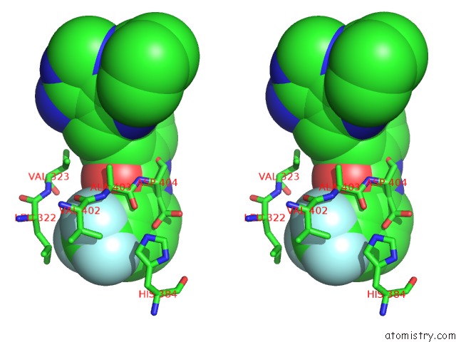
Stereo pair view
Mono view
Stereo pair view
A full contact list of Fluorine with other atoms in the F binding
site number 1 of Crystal Structure of C-Src in Complex with Pyrazolopyrimidine 3 within 5.0Å range:
|
Fluorine binding site 2 out of 3 in 3el7
Go back to
Fluorine binding site 2 out
of 3 in the Crystal Structure of C-Src in Complex with Pyrazolopyrimidine 3
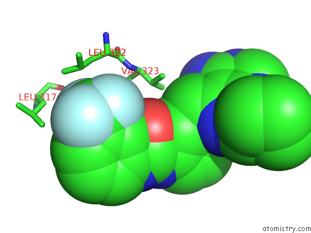
Mono view
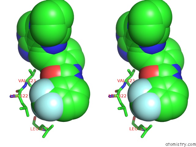
Stereo pair view
Mono view
Stereo pair view
A full contact list of Fluorine with other atoms in the F binding
site number 2 of Crystal Structure of C-Src in Complex with Pyrazolopyrimidine 3 within 5.0Å range:
|
Fluorine binding site 3 out of 3 in 3el7
Go back to
Fluorine binding site 3 out
of 3 in the Crystal Structure of C-Src in Complex with Pyrazolopyrimidine 3
Mono view
Stereo pair view
Mono view
Stereo pair view
A full contact list of Fluorine with other atoms in the F binding
site number 3 of Crystal Structure of C-Src in Complex with Pyrazolopyrimidine 3 within 5.0Å range:
|
Reference:
A.C.Dar,
M.S.Lopez,
K.M.Shokat.
Small Molecule Recognition of C-Src Via the Imatinib-Binding Conformation. Chem.Biol. V. 15 1015 2008.
ISSN: ISSN 1074-5521
PubMed: 18940662
DOI: 10.1016/J.CHEMBIOL.2008.09.007
Page generated: Wed Jul 31 18:18:30 2024
ISSN: ISSN 1074-5521
PubMed: 18940662
DOI: 10.1016/J.CHEMBIOL.2008.09.007
Last articles
Zn in 9J0NZn in 9J0O
Zn in 9J0P
Zn in 9FJX
Zn in 9EKB
Zn in 9C0F
Zn in 9CAH
Zn in 9CH0
Zn in 9CH3
Zn in 9CH1