Fluorine »
PDB 3fls-3g70 »
3fqs »
Fluorine in PDB 3fqs: Crystal Structure of Spleen Tyrosine Kinase Complexed with R406
Enzymatic activity of Crystal Structure of Spleen Tyrosine Kinase Complexed with R406
All present enzymatic activity of Crystal Structure of Spleen Tyrosine Kinase Complexed with R406:
2.7.10.2;
2.7.10.2;
Protein crystallography data
The structure of Crystal Structure of Spleen Tyrosine Kinase Complexed with R406, PDB code: 3fqs
was solved by
A.Kuglstatter,
A.G.Villasenor,
with X-Ray Crystallography technique. A brief refinement statistics is given in the table below:
| Resolution Low / High (Å) | 42.52 / 2.10 |
| Space group | P 21 21 21 |
| Cell size a, b, c (Å), α, β, γ (°) | 39.971, 85.079, 91.144, 90.00, 90.00, 90.00 |
| R / Rfree (%) | 21.6 / 25.3 |
Fluorine Binding Sites:
The binding sites of Fluorine atom in the Crystal Structure of Spleen Tyrosine Kinase Complexed with R406
(pdb code 3fqs). This binding sites where shown within
5.0 Angstroms radius around Fluorine atom.
In total only one binding site of Fluorine was determined in the Crystal Structure of Spleen Tyrosine Kinase Complexed with R406, PDB code: 3fqs:
In total only one binding site of Fluorine was determined in the Crystal Structure of Spleen Tyrosine Kinase Complexed with R406, PDB code: 3fqs:
Fluorine binding site 1 out of 1 in 3fqs
Go back to
Fluorine binding site 1 out
of 1 in the Crystal Structure of Spleen Tyrosine Kinase Complexed with R406

Mono view
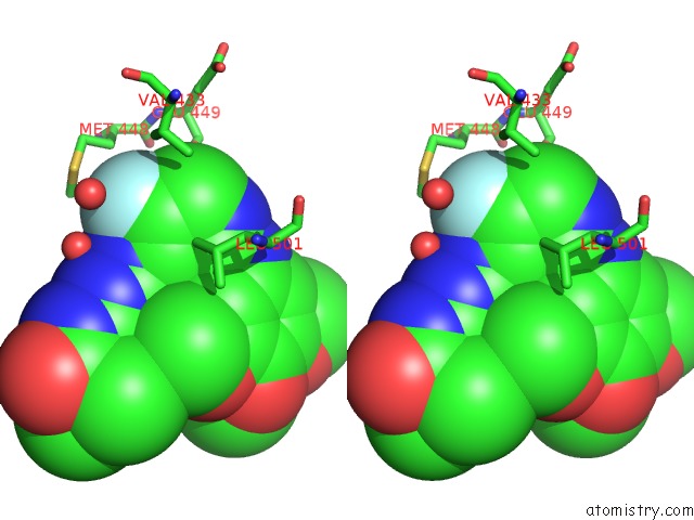
Stereo pair view
Mono view
Stereo pair view
A full contact list of Fluorine with other atoms in the F binding
site number 1 of Crystal Structure of Spleen Tyrosine Kinase Complexed with R406 within 5.0Å range:
|
Reference:
A.G.Villasenor,
R.Kondru,
H.Ho,
S.Wang,
E.Papp,
D.Shaw,
J.W.Barnett,
M.F.Browner,
A.Kuglstatter.
Structural Insights For Design of Potent Spleen Tyrosine Kinase Inhibitors From Crystallographic Analysis of Three Inhibitor Complexes. Chem.Biol.Drug Des. V. 73 466 2009.
ISSN: ISSN 1747-0277
PubMed: 19220318
DOI: 10.1111/J.1747-0285.2009.00785.X
Page generated: Wed Jul 31 18:40:27 2024
ISSN: ISSN 1747-0277
PubMed: 19220318
DOI: 10.1111/J.1747-0285.2009.00785.X
Last articles
Zn in 9MJ5Zn in 9HNW
Zn in 9G0L
Zn in 9FNE
Zn in 9DZN
Zn in 9E0I
Zn in 9D32
Zn in 9DAK
Zn in 8ZXC
Zn in 8ZUF