Fluorine »
PDB 3fls-3g70 »
3fuc »
Fluorine in PDB 3fuc: Recombinant Calf Purine Nucleoside Phosphorylase in A Binary Complex with Multisubstrate Analogue Inhibitor 9-(5',5'-Difluoro-5'- Phosphonopentyl)-9-Deazaguanine Structure in A New Space Group with One Full Trimer in the Asymmetric Unit
Enzymatic activity of Recombinant Calf Purine Nucleoside Phosphorylase in A Binary Complex with Multisubstrate Analogue Inhibitor 9-(5',5'-Difluoro-5'- Phosphonopentyl)-9-Deazaguanine Structure in A New Space Group with One Full Trimer in the Asymmetric Unit
All present enzymatic activity of Recombinant Calf Purine Nucleoside Phosphorylase in A Binary Complex with Multisubstrate Analogue Inhibitor 9-(5',5'-Difluoro-5'- Phosphonopentyl)-9-Deazaguanine Structure in A New Space Group with One Full Trimer in the Asymmetric Unit:
2.4.2.1;
2.4.2.1;
Protein crystallography data
The structure of Recombinant Calf Purine Nucleoside Phosphorylase in A Binary Complex with Multisubstrate Analogue Inhibitor 9-(5',5'-Difluoro-5'- Phosphonopentyl)-9-Deazaguanine Structure in A New Space Group with One Full Trimer in the Asymmetric Unit, PDB code: 3fuc
was solved by
M.Bochtler,
K.Breer,
A.Bzowska,
G.Chojnowski,
M.Hashimoto,
S.Hikishima,
M.Narczyk,
B.Wielgus-Kutrowska,
T.Yokomatsu,
with X-Ray Crystallography technique. A brief refinement statistics is given in the table below:
| Resolution Low / High (Å) | 10.00 / 1.45 |
| Space group | C 1 2 1 |
| Cell size a, b, c (Å), α, β, γ (°) | 135.795, 78.253, 94.901, 90.00, 97.19, 90.00 |
| R / Rfree (%) | 18 / 19.9 |
Other elements in 3fuc:
The structure of Recombinant Calf Purine Nucleoside Phosphorylase in A Binary Complex with Multisubstrate Analogue Inhibitor 9-(5',5'-Difluoro-5'- Phosphonopentyl)-9-Deazaguanine Structure in A New Space Group with One Full Trimer in the Asymmetric Unit also contains other interesting chemical elements:
| Magnesium | (Mg) | 3 atoms |
Fluorine Binding Sites:
The binding sites of Fluorine atom in the Recombinant Calf Purine Nucleoside Phosphorylase in A Binary Complex with Multisubstrate Analogue Inhibitor 9-(5',5'-Difluoro-5'- Phosphonopentyl)-9-Deazaguanine Structure in A New Space Group with One Full Trimer in the Asymmetric Unit
(pdb code 3fuc). This binding sites where shown within
5.0 Angstroms radius around Fluorine atom.
In total 6 binding sites of Fluorine where determined in the Recombinant Calf Purine Nucleoside Phosphorylase in A Binary Complex with Multisubstrate Analogue Inhibitor 9-(5',5'-Difluoro-5'- Phosphonopentyl)-9-Deazaguanine Structure in A New Space Group with One Full Trimer in the Asymmetric Unit, PDB code: 3fuc:
Jump to Fluorine binding site number: 1; 2; 3; 4; 5; 6;
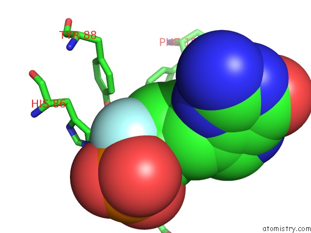
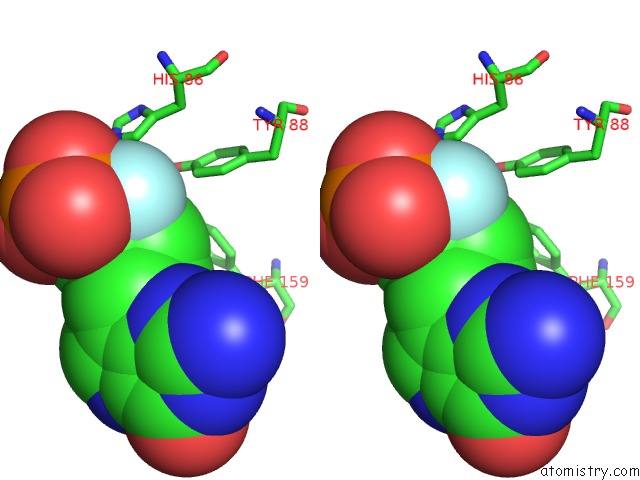
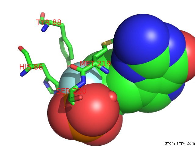
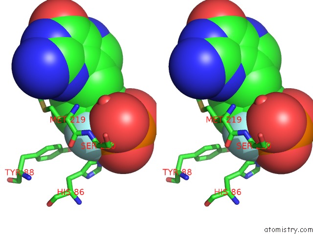
Fluorine binding site 1 out of 6 in 3fuc
Go back to
Fluorine binding site 1 out
of 6 in the Recombinant Calf Purine Nucleoside Phosphorylase in A Binary Complex with Multisubstrate Analogue Inhibitor 9-(5',5'-Difluoro-5'- Phosphonopentyl)-9-Deazaguanine Structure in A New Space Group with One Full Trimer in the Asymmetric Unit
Mono view

Stereo pair view
Mono view
Stereo pair view
A full contact list of Fluorine with other atoms in the F binding
site number 1 of Recombinant Calf Purine Nucleoside Phosphorylase in A Binary Complex with Multisubstrate Analogue Inhibitor 9-(5',5'-Difluoro-5'- Phosphonopentyl)-9-Deazaguanine Structure in A New Space Group with One Full Trimer in the Asymmetric Unit within 5.0Å range:
|
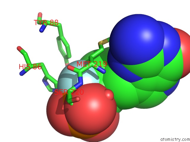
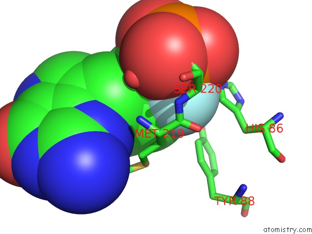
Fluorine binding site 2 out of 6 in 3fuc
Go back to
Fluorine binding site 2 out
of 6 in the Recombinant Calf Purine Nucleoside Phosphorylase in A Binary Complex with Multisubstrate Analogue Inhibitor 9-(5',5'-Difluoro-5'- Phosphonopentyl)-9-Deazaguanine Structure in A New Space Group with One Full Trimer in the Asymmetric Unit
Mono view
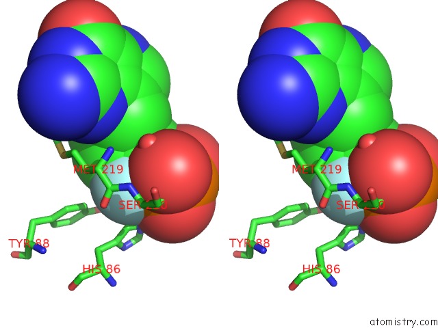
Stereo pair view
Mono view
Stereo pair view
A full contact list of Fluorine with other atoms in the F binding
site number 2 of Recombinant Calf Purine Nucleoside Phosphorylase in A Binary Complex with Multisubstrate Analogue Inhibitor 9-(5',5'-Difluoro-5'- Phosphonopentyl)-9-Deazaguanine Structure in A New Space Group with One Full Trimer in the Asymmetric Unit within 5.0Å range:
|
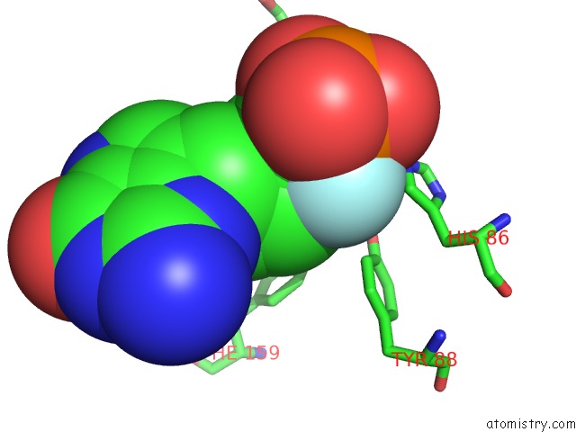
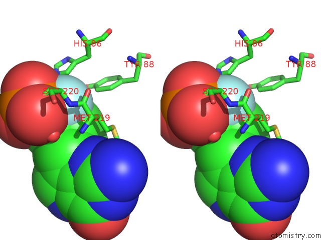
Fluorine binding site 3 out of 6 in 3fuc
Go back to
Fluorine binding site 3 out
of 6 in the Recombinant Calf Purine Nucleoside Phosphorylase in A Binary Complex with Multisubstrate Analogue Inhibitor 9-(5',5'-Difluoro-5'- Phosphonopentyl)-9-Deazaguanine Structure in A New Space Group with One Full Trimer in the Asymmetric Unit
Mono view
Stereo pair view
Mono view
Stereo pair view
A full contact list of Fluorine with other atoms in the F binding
site number 3 of Recombinant Calf Purine Nucleoside Phosphorylase in A Binary Complex with Multisubstrate Analogue Inhibitor 9-(5',5'-Difluoro-5'- Phosphonopentyl)-9-Deazaguanine Structure in A New Space Group with One Full Trimer in the Asymmetric Unit within 5.0Å range:
|
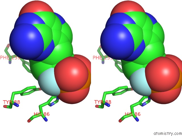
Fluorine binding site 4 out of 6 in 3fuc
Go back to
Fluorine binding site 4 out
of 6 in the Recombinant Calf Purine Nucleoside Phosphorylase in A Binary Complex with Multisubstrate Analogue Inhibitor 9-(5',5'-Difluoro-5'- Phosphonopentyl)-9-Deazaguanine Structure in A New Space Group with One Full Trimer in the Asymmetric Unit
Mono view
Stereo pair view
Mono view
Stereo pair view
A full contact list of Fluorine with other atoms in the F binding
site number 4 of Recombinant Calf Purine Nucleoside Phosphorylase in A Binary Complex with Multisubstrate Analogue Inhibitor 9-(5',5'-Difluoro-5'- Phosphonopentyl)-9-Deazaguanine Structure in A New Space Group with One Full Trimer in the Asymmetric Unit within 5.0Å range:
|
Fluorine binding site 5 out of 6 in 3fuc
Go back to
Fluorine binding site 5 out
of 6 in the Recombinant Calf Purine Nucleoside Phosphorylase in A Binary Complex with Multisubstrate Analogue Inhibitor 9-(5',5'-Difluoro-5'- Phosphonopentyl)-9-Deazaguanine Structure in A New Space Group with One Full Trimer in the Asymmetric Unit

Mono view
Stereo pair view
Mono view
Stereo pair view
A full contact list of Fluorine with other atoms in the F binding
site number 5 of Recombinant Calf Purine Nucleoside Phosphorylase in A Binary Complex with Multisubstrate Analogue Inhibitor 9-(5',5'-Difluoro-5'- Phosphonopentyl)-9-Deazaguanine Structure in A New Space Group with One Full Trimer in the Asymmetric Unit within 5.0Å range:
|
Fluorine binding site 6 out of 6 in 3fuc
Go back to
Fluorine binding site 6 out
of 6 in the Recombinant Calf Purine Nucleoside Phosphorylase in A Binary Complex with Multisubstrate Analogue Inhibitor 9-(5',5'-Difluoro-5'- Phosphonopentyl)-9-Deazaguanine Structure in A New Space Group with One Full Trimer in the Asymmetric Unit
Mono view
Stereo pair view
Mono view
Stereo pair view
A full contact list of Fluorine with other atoms in the F binding
site number 6 of Recombinant Calf Purine Nucleoside Phosphorylase in A Binary Complex with Multisubstrate Analogue Inhibitor 9-(5',5'-Difluoro-5'- Phosphonopentyl)-9-Deazaguanine Structure in A New Space Group with One Full Trimer in the Asymmetric Unit within 5.0Å range:
|
Reference:
G.Chojnowski,
K.Breer,
M.Narczyk,
B.Wielgus-Kutrowska,
H.Czapinska,
M.Hashimoto,
S.Hikishima,
T.Yokomatsu,
M.Bochtler,
A.Girstun,
K.Staron,
A.Bzowska.
1.45 A Resolution Crystal Structure of Recombinant Pnp in Complex with A Pm Multisubstrate Analogue Inhibitor Bearing One Feature of the Postulated Transition State. Biochem.Biophys.Res.Commun. V. 391 703 2010.
ISSN: ISSN 0006-291X
PubMed: 19944078
DOI: 10.1016/J.BBRC.2009.11.124
Page generated: Wed Jul 31 18:41:29 2024
ISSN: ISSN 0006-291X
PubMed: 19944078
DOI: 10.1016/J.BBRC.2009.11.124
Last articles
Zn in 9MJ5Zn in 9HNW
Zn in 9G0L
Zn in 9FNE
Zn in 9DZN
Zn in 9E0I
Zn in 9D32
Zn in 9DAK
Zn in 8ZXC
Zn in 8ZUF