Fluorine »
PDB 3g72-3gwv »
3gc9 »
Fluorine in PDB 3gc9: The Structure of P38BETA C119S, C162S in Complex with A Dihydroquinazolinone Inhibitor
Enzymatic activity of The Structure of P38BETA C119S, C162S in Complex with A Dihydroquinazolinone Inhibitor
All present enzymatic activity of The Structure of P38BETA C119S, C162S in Complex with A Dihydroquinazolinone Inhibitor:
2.7.11.24;
2.7.11.24;
Protein crystallography data
The structure of The Structure of P38BETA C119S, C162S in Complex with A Dihydroquinazolinone Inhibitor, PDB code: 3gc9
was solved by
G.Scapin,
S.B.Patel,
with X-Ray Crystallography technique. A brief refinement statistics is given in the table below:
| Resolution Low / High (Å) | 30.00 / 2.05 |
| Space group | P 1 21 1 |
| Cell size a, b, c (Å), α, β, γ (°) | 39.184, 158.823, 60.874, 90.00, 91.59, 90.00 |
| R / Rfree (%) | 22.2 / 27.5 |
Other elements in 3gc9:
The structure of The Structure of P38BETA C119S, C162S in Complex with A Dihydroquinazolinone Inhibitor also contains other interesting chemical elements:
| Zinc | (Zn) | 1 atom |
| Chlorine | (Cl) | 6 atoms |
| Sodium | (Na) | 2 atoms |
Fluorine Binding Sites:
The binding sites of Fluorine atom in the The Structure of P38BETA C119S, C162S in Complex with A Dihydroquinazolinone Inhibitor
(pdb code 3gc9). This binding sites where shown within
5.0 Angstroms radius around Fluorine atom.
In total 2 binding sites of Fluorine where determined in the The Structure of P38BETA C119S, C162S in Complex with A Dihydroquinazolinone Inhibitor, PDB code: 3gc9:
Jump to Fluorine binding site number: 1; 2;
In total 2 binding sites of Fluorine where determined in the The Structure of P38BETA C119S, C162S in Complex with A Dihydroquinazolinone Inhibitor, PDB code: 3gc9:
Jump to Fluorine binding site number: 1; 2;
Fluorine binding site 1 out of 2 in 3gc9
Go back to
Fluorine binding site 1 out
of 2 in the The Structure of P38BETA C119S, C162S in Complex with A Dihydroquinazolinone Inhibitor
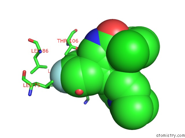
Mono view
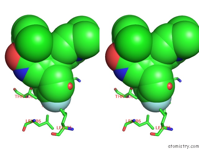
Stereo pair view
Mono view
Stereo pair view
A full contact list of Fluorine with other atoms in the F binding
site number 1 of The Structure of P38BETA C119S, C162S in Complex with A Dihydroquinazolinone Inhibitor within 5.0Å range:
|
Fluorine binding site 2 out of 2 in 3gc9
Go back to
Fluorine binding site 2 out
of 2 in the The Structure of P38BETA C119S, C162S in Complex with A Dihydroquinazolinone Inhibitor
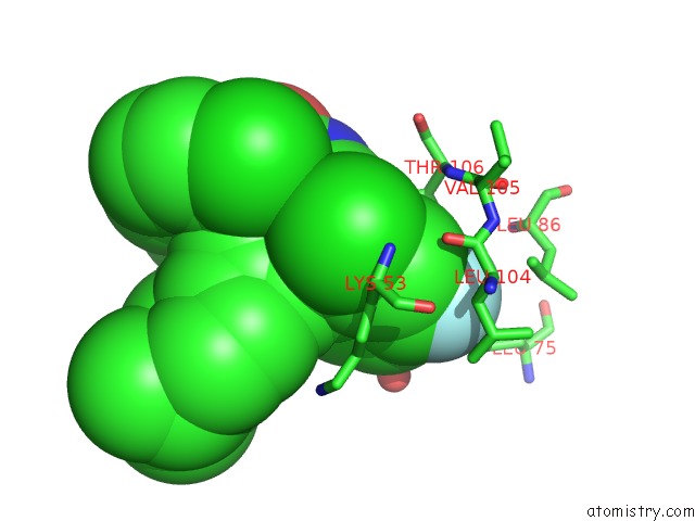
Mono view
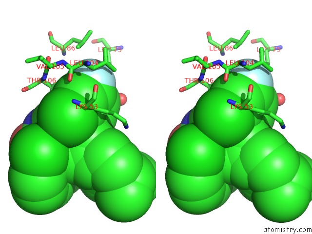
Stereo pair view
Mono view
Stereo pair view
A full contact list of Fluorine with other atoms in the F binding
site number 2 of The Structure of P38BETA C119S, C162S in Complex with A Dihydroquinazolinone Inhibitor within 5.0Å range:
|
Reference:
S.B.Patel,
P.M.Cameron,
S.J.O'keefe,
B.Frantz-Wattley,
J.Thompson,
E.A.O'neill,
T.Tennis,
L.Liu,
J.W.Becker,
G.Scapin.
The Three-Dimensional Structure of Map Kinase P38BETA: Different Features of the Atp-Binding Site in P38BETA Compared with P38ALPHA. Acta Crystallogr.,Sect.D V. 65 777 2009.
ISSN: ISSN 0907-4449
PubMed: 19622861
DOI: 10.1107/S090744490901600X
Page generated: Mon Jul 14 16:32:05 2025
ISSN: ISSN 0907-4449
PubMed: 19622861
DOI: 10.1107/S090744490901600X
Last articles
Fe in 2YXOFe in 2YRS
Fe in 2YXC
Fe in 2YNM
Fe in 2YVJ
Fe in 2YP1
Fe in 2YU2
Fe in 2YU1
Fe in 2YQB
Fe in 2YOO