Fluorine »
PDB 3g72-3gwv »
3gn8 »
Fluorine in PDB 3gn8: X-Ray Crystal Structure of ANCGR2 in Complex with Dexamethasone
Protein crystallography data
The structure of X-Ray Crystal Structure of ANCGR2 in Complex with Dexamethasone, PDB code: 3gn8
was solved by
E.A.Ortlund,
with X-Ray Crystallography technique. A brief refinement statistics is given in the table below:
| Resolution Low / High (Å) | 49.01 / 2.50 |
| Space group | P 61 |
| Cell size a, b, c (Å), α, β, γ (°) | 104.218, 104.218, 144.166, 90.00, 90.00, 120.00 |
| R / Rfree (%) | 19.6 / 25.5 |
Fluorine Binding Sites:
The binding sites of Fluorine atom in the X-Ray Crystal Structure of ANCGR2 in Complex with Dexamethasone
(pdb code 3gn8). This binding sites where shown within
5.0 Angstroms radius around Fluorine atom.
In total 2 binding sites of Fluorine where determined in the X-Ray Crystal Structure of ANCGR2 in Complex with Dexamethasone, PDB code: 3gn8:
Jump to Fluorine binding site number: 1; 2;
In total 2 binding sites of Fluorine where determined in the X-Ray Crystal Structure of ANCGR2 in Complex with Dexamethasone, PDB code: 3gn8:
Jump to Fluorine binding site number: 1; 2;
Fluorine binding site 1 out of 2 in 3gn8
Go back to
Fluorine binding site 1 out
of 2 in the X-Ray Crystal Structure of ANCGR2 in Complex with Dexamethasone
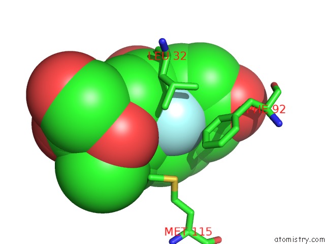
Mono view
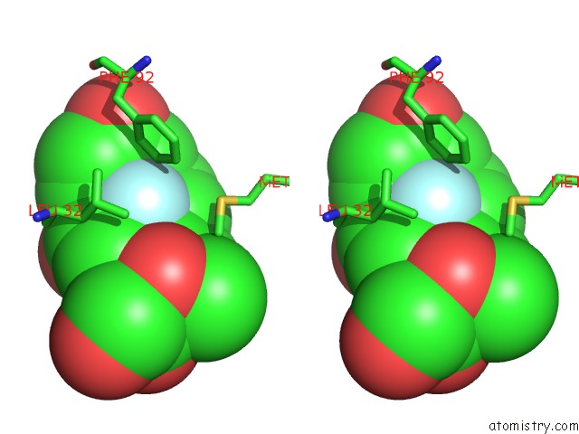
Stereo pair view
Mono view
Stereo pair view
A full contact list of Fluorine with other atoms in the F binding
site number 1 of X-Ray Crystal Structure of ANCGR2 in Complex with Dexamethasone within 5.0Å range:
|
Fluorine binding site 2 out of 2 in 3gn8
Go back to
Fluorine binding site 2 out
of 2 in the X-Ray Crystal Structure of ANCGR2 in Complex with Dexamethasone
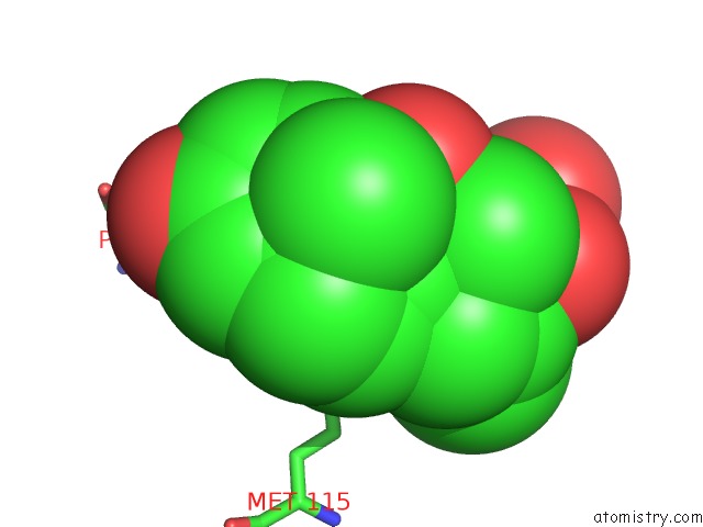
Mono view
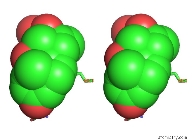
Stereo pair view
Mono view
Stereo pair view
A full contact list of Fluorine with other atoms in the F binding
site number 2 of X-Ray Crystal Structure of ANCGR2 in Complex with Dexamethasone within 5.0Å range:
|
Reference:
J.T.Bridgham,
E.A.Ortlund,
J.W.Thornton.
An Epistatic Ratchet Constrains the Direction of Glucocorticoid Receptor Evolution Nature V. 461 515 2009.
ISSN: ISSN 0028-0836
PubMed: 19779450
DOI: 10.1038/NATURE08249
Page generated: Wed Jul 31 18:56:01 2024
ISSN: ISSN 0028-0836
PubMed: 19779450
DOI: 10.1038/NATURE08249
Last articles
Zn in 9J0NZn in 9J0O
Zn in 9J0P
Zn in 9FJX
Zn in 9EKB
Zn in 9C0F
Zn in 9CAH
Zn in 9CH0
Zn in 9CH3
Zn in 9CH1