Fluorine »
PDB 3g72-3gwv »
3gpt »
Fluorine in PDB 3gpt: Crystal Structure of the Yeast 20S Proteasome in Complex with Salinosporamide Derivatives: Slow Substrate Ligand
Enzymatic activity of Crystal Structure of the Yeast 20S Proteasome in Complex with Salinosporamide Derivatives: Slow Substrate Ligand
All present enzymatic activity of Crystal Structure of the Yeast 20S Proteasome in Complex with Salinosporamide Derivatives: Slow Substrate Ligand:
3.4.25.1;
3.4.25.1;
Protein crystallography data
The structure of Crystal Structure of the Yeast 20S Proteasome in Complex with Salinosporamide Derivatives: Slow Substrate Ligand, PDB code: 3gpt
was solved by
M.Groll,
V.R.Macherla,
R.R.Manam,
K.A.M.Arthur,
C.B.Potts,
with X-Ray Crystallography technique. A brief refinement statistics is given in the table below:
| Resolution Low / High (Å) | 15.00 / 2.41 |
| Space group | P 1 21 1 |
| Cell size a, b, c (Å), α, β, γ (°) | 134.000, 301.260, 143.810, 90.00, 112.56, 90.00 |
| R / Rfree (%) | 22.6 / 24.5 |
Fluorine Binding Sites:
The binding sites of Fluorine atom in the Crystal Structure of the Yeast 20S Proteasome in Complex with Salinosporamide Derivatives: Slow Substrate Ligand
(pdb code 3gpt). This binding sites where shown within
5.0 Angstroms radius around Fluorine atom.
In total 6 binding sites of Fluorine where determined in the Crystal Structure of the Yeast 20S Proteasome in Complex with Salinosporamide Derivatives: Slow Substrate Ligand, PDB code: 3gpt:
Jump to Fluorine binding site number: 1; 2; 3; 4; 5; 6;
In total 6 binding sites of Fluorine where determined in the Crystal Structure of the Yeast 20S Proteasome in Complex with Salinosporamide Derivatives: Slow Substrate Ligand, PDB code: 3gpt:
Jump to Fluorine binding site number: 1; 2; 3; 4; 5; 6;
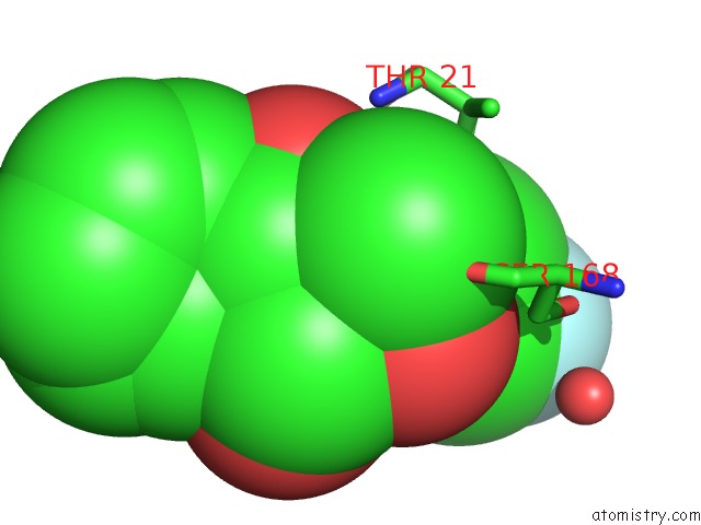
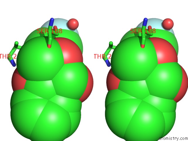
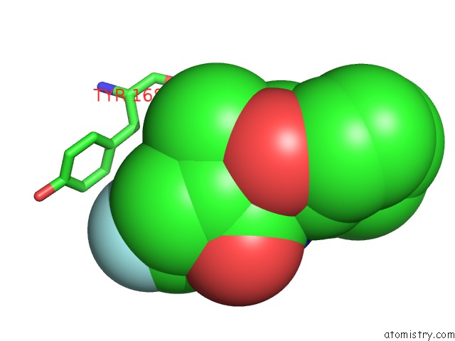
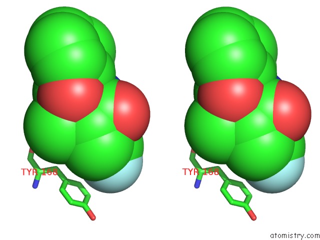
Fluorine binding site 1 out of 6 in 3gpt
Go back to
Fluorine binding site 1 out
of 6 in the Crystal Structure of the Yeast 20S Proteasome in Complex with Salinosporamide Derivatives: Slow Substrate Ligand
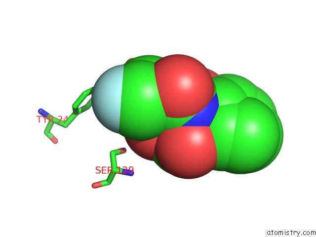
Mono view
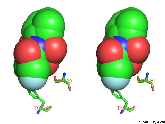
Stereo pair view
Mono view
Stereo pair view
A full contact list of Fluorine with other atoms in the F binding
site number 1 of Crystal Structure of the Yeast 20S Proteasome in Complex with Salinosporamide Derivatives: Slow Substrate Ligand within 5.0Å range:
|
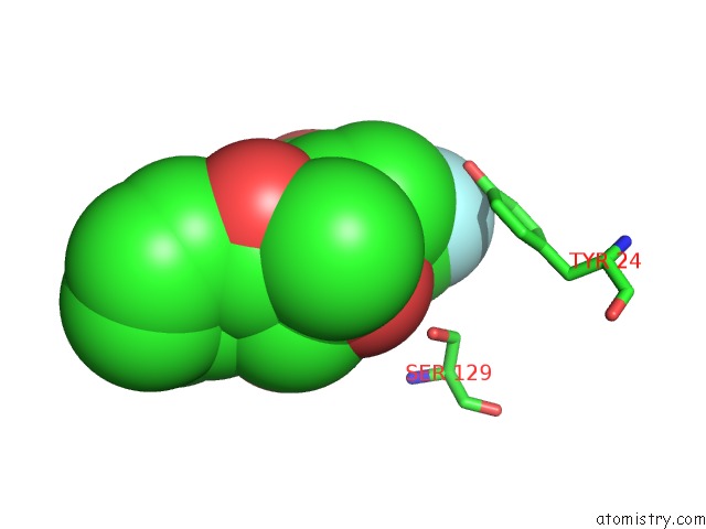
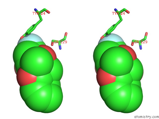
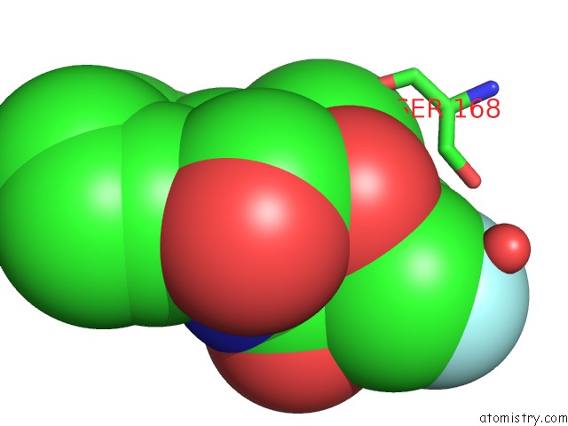
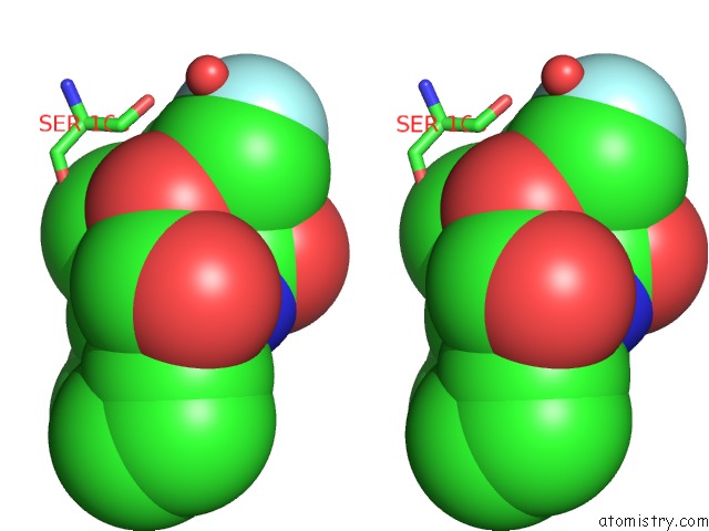
Fluorine binding site 2 out of 6 in 3gpt
Go back to
Fluorine binding site 2 out
of 6 in the Crystal Structure of the Yeast 20S Proteasome in Complex with Salinosporamide Derivatives: Slow Substrate Ligand
Mono view
Stereo pair view
Mono view
Stereo pair view
A full contact list of Fluorine with other atoms in the F binding
site number 2 of Crystal Structure of the Yeast 20S Proteasome in Complex with Salinosporamide Derivatives: Slow Substrate Ligand within 5.0Å range:
|
Fluorine binding site 3 out of 6 in 3gpt
Go back to
Fluorine binding site 3 out
of 6 in the Crystal Structure of the Yeast 20S Proteasome in Complex with Salinosporamide Derivatives: Slow Substrate Ligand
Mono view
Stereo pair view
Mono view
Stereo pair view
A full contact list of Fluorine with other atoms in the F binding
site number 3 of Crystal Structure of the Yeast 20S Proteasome in Complex with Salinosporamide Derivatives: Slow Substrate Ligand within 5.0Å range:
|
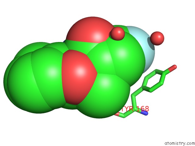
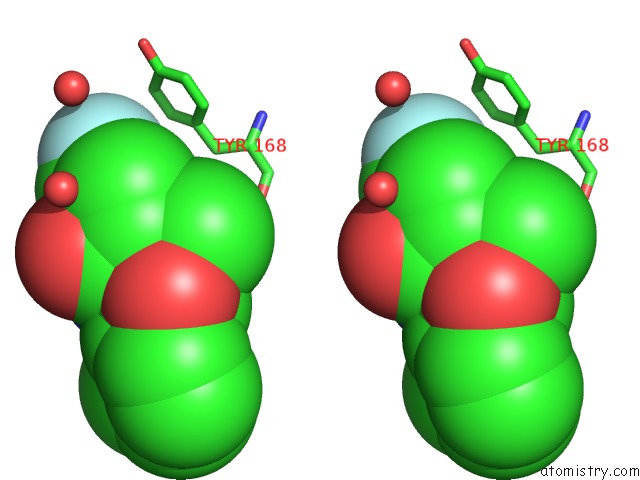
Fluorine binding site 4 out of 6 in 3gpt
Go back to
Fluorine binding site 4 out
of 6 in the Crystal Structure of the Yeast 20S Proteasome in Complex with Salinosporamide Derivatives: Slow Substrate Ligand
Mono view
Stereo pair view
Mono view
Stereo pair view
A full contact list of Fluorine with other atoms in the F binding
site number 4 of Crystal Structure of the Yeast 20S Proteasome in Complex with Salinosporamide Derivatives: Slow Substrate Ligand within 5.0Å range:
|
Fluorine binding site 5 out of 6 in 3gpt
Go back to
Fluorine binding site 5 out
of 6 in the Crystal Structure of the Yeast 20S Proteasome in Complex with Salinosporamide Derivatives: Slow Substrate Ligand
Mono view
Stereo pair view
Mono view
Stereo pair view
A full contact list of Fluorine with other atoms in the F binding
site number 5 of Crystal Structure of the Yeast 20S Proteasome in Complex with Salinosporamide Derivatives: Slow Substrate Ligand within 5.0Å range:
|
Fluorine binding site 6 out of 6 in 3gpt
Go back to
Fluorine binding site 6 out
of 6 in the Crystal Structure of the Yeast 20S Proteasome in Complex with Salinosporamide Derivatives: Slow Substrate Ligand
Mono view
Stereo pair view
Mono view
Stereo pair view
A full contact list of Fluorine with other atoms in the F binding
site number 6 of Crystal Structure of the Yeast 20S Proteasome in Complex with Salinosporamide Derivatives: Slow Substrate Ligand within 5.0Å range:
|
Reference:
M.Groll,
K.A.Mcarthur,
V.R.Macherla,
R.R.Manam,
B.C.Potts.
Snapshots of the Fluorosalinosporamide/20S Complex Offer Mechanistic Insights For Fine Tuning Proteasome Inhibition J.Med.Chem. V. 52 5420 2009.
ISSN: ISSN 0022-2623
PubMed: 19678642
DOI: 10.1021/JM900559X
Page generated: Wed Jul 31 18:58:13 2024
ISSN: ISSN 0022-2623
PubMed: 19678642
DOI: 10.1021/JM900559X
Last articles
Zn in 9MJ5Zn in 9HNW
Zn in 9G0L
Zn in 9FNE
Zn in 9DZN
Zn in 9E0I
Zn in 9D32
Zn in 9DAK
Zn in 8ZXC
Zn in 8ZUF