Fluorine »
PDB 3g72-3gwv »
3gql »
Fluorine in PDB 3gql: Crystal Structure of Activated Receptor Tyrosine Kinase in Complex with Substrates
Enzymatic activity of Crystal Structure of Activated Receptor Tyrosine Kinase in Complex with Substrates
All present enzymatic activity of Crystal Structure of Activated Receptor Tyrosine Kinase in Complex with Substrates:
2.7.10.1;
2.7.10.1;
Protein crystallography data
The structure of Crystal Structure of Activated Receptor Tyrosine Kinase in Complex with Substrates, PDB code: 3gql
was solved by
J.H.Bae,
E.D.Lew,
S.Yuzawa,
F.Tome,
I.Lax,
J.Schlessinger,
with X-Ray Crystallography technique. A brief refinement statistics is given in the table below:
| Resolution Low / High (Å) | 36.01 / 2.80 |
| Space group | C 1 2 1 |
| Cell size a, b, c (Å), α, β, γ (°) | 194.168, 78.265, 98.458, 90.00, 110.55, 90.00 |
| R / Rfree (%) | 25.2 / 28.9 |
Fluorine Binding Sites:
The binding sites of Fluorine atom in the Crystal Structure of Activated Receptor Tyrosine Kinase in Complex with Substrates
(pdb code 3gql). This binding sites where shown within
5.0 Angstroms radius around Fluorine atom.
In total 6 binding sites of Fluorine where determined in the Crystal Structure of Activated Receptor Tyrosine Kinase in Complex with Substrates, PDB code: 3gql:
Jump to Fluorine binding site number: 1; 2; 3; 4; 5; 6;
In total 6 binding sites of Fluorine where determined in the Crystal Structure of Activated Receptor Tyrosine Kinase in Complex with Substrates, PDB code: 3gql:
Jump to Fluorine binding site number: 1; 2; 3; 4; 5; 6;
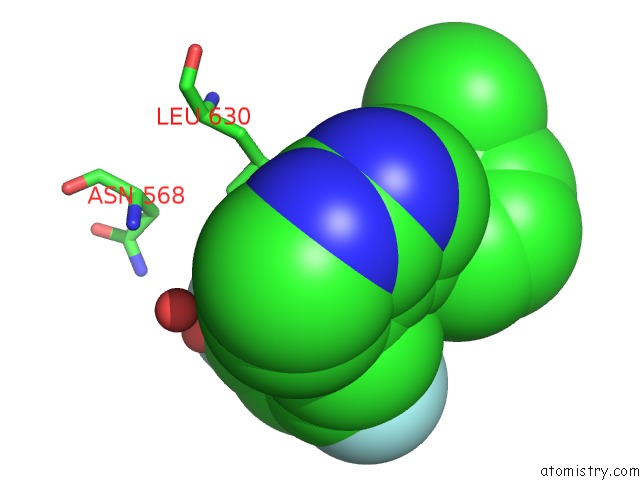
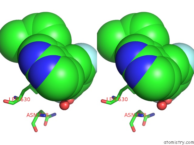
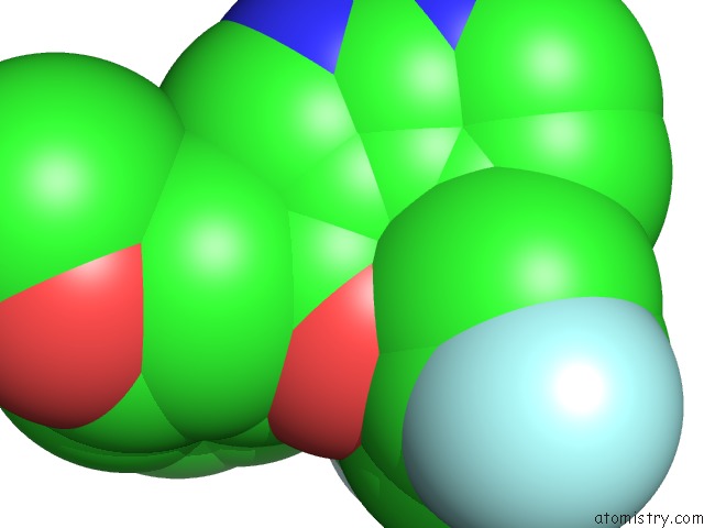
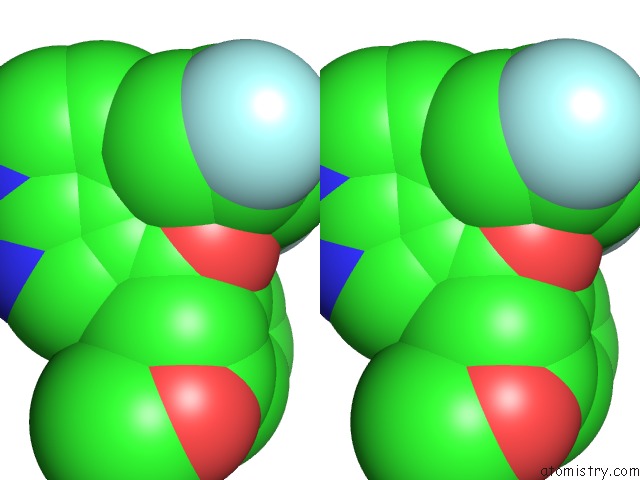
Fluorine binding site 1 out of 6 in 3gql
Go back to
Fluorine binding site 1 out
of 6 in the Crystal Structure of Activated Receptor Tyrosine Kinase in Complex with Substrates
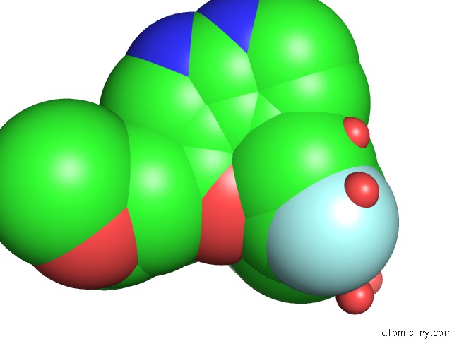
Mono view
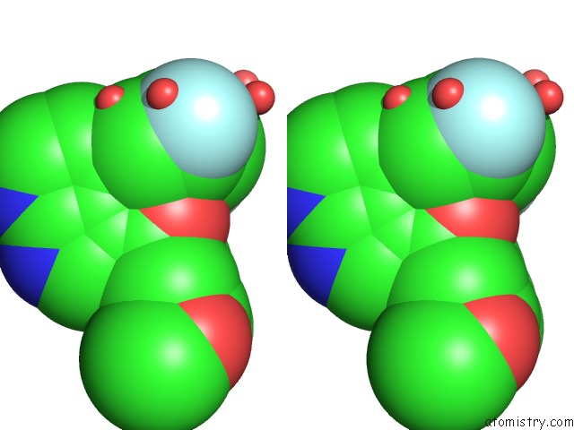
Stereo pair view
Mono view
Stereo pair view
A full contact list of Fluorine with other atoms in the F binding
site number 1 of Crystal Structure of Activated Receptor Tyrosine Kinase in Complex with Substrates within 5.0Å range:
|
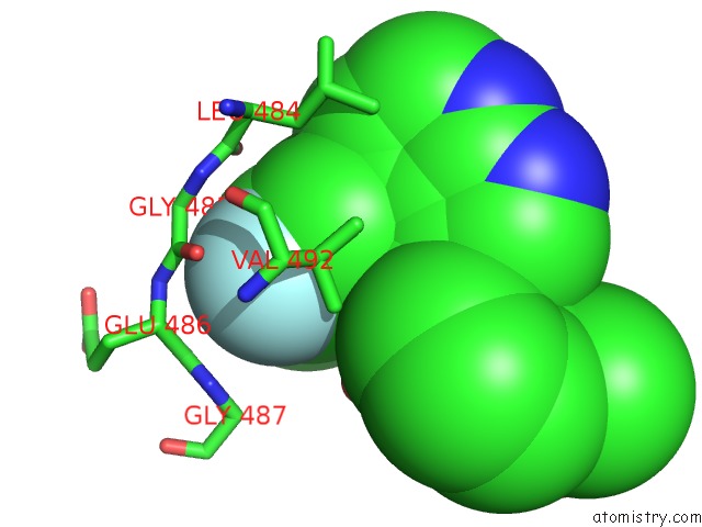
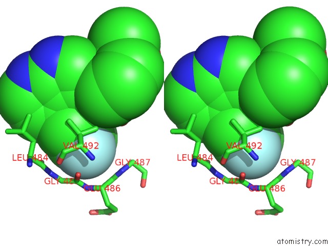
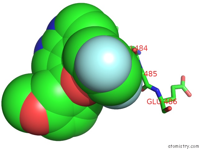
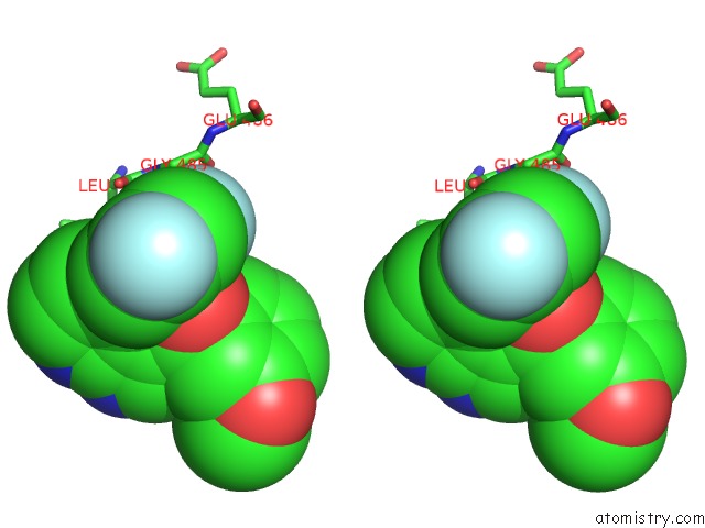
Fluorine binding site 2 out of 6 in 3gql
Go back to
Fluorine binding site 2 out
of 6 in the Crystal Structure of Activated Receptor Tyrosine Kinase in Complex with Substrates
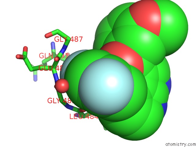
Mono view
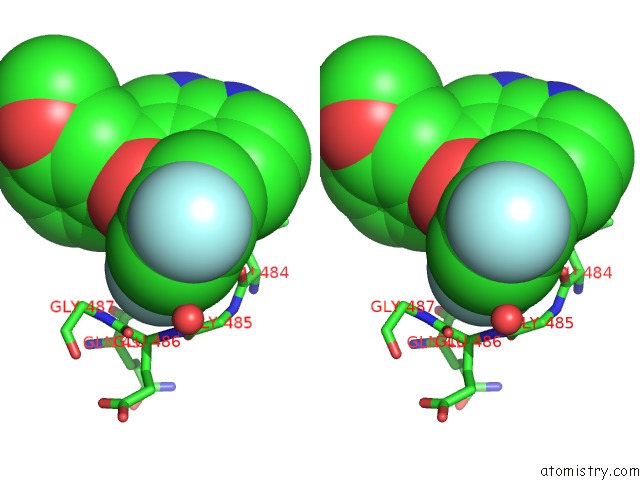
Stereo pair view
Mono view
Stereo pair view
A full contact list of Fluorine with other atoms in the F binding
site number 2 of Crystal Structure of Activated Receptor Tyrosine Kinase in Complex with Substrates within 5.0Å range:
|
Fluorine binding site 3 out of 6 in 3gql
Go back to
Fluorine binding site 3 out
of 6 in the Crystal Structure of Activated Receptor Tyrosine Kinase in Complex with Substrates
Mono view
Stereo pair view
Mono view
Stereo pair view
A full contact list of Fluorine with other atoms in the F binding
site number 3 of Crystal Structure of Activated Receptor Tyrosine Kinase in Complex with Substrates within 5.0Å range:
|
Fluorine binding site 4 out of 6 in 3gql
Go back to
Fluorine binding site 4 out
of 6 in the Crystal Structure of Activated Receptor Tyrosine Kinase in Complex with Substrates
Mono view
Stereo pair view
Mono view
Stereo pair view
A full contact list of Fluorine with other atoms in the F binding
site number 4 of Crystal Structure of Activated Receptor Tyrosine Kinase in Complex with Substrates within 5.0Å range:
|
Fluorine binding site 5 out of 6 in 3gql
Go back to
Fluorine binding site 5 out
of 6 in the Crystal Structure of Activated Receptor Tyrosine Kinase in Complex with Substrates
Mono view
Stereo pair view
Mono view
Stereo pair view
A full contact list of Fluorine with other atoms in the F binding
site number 5 of Crystal Structure of Activated Receptor Tyrosine Kinase in Complex with Substrates within 5.0Å range:
|
Fluorine binding site 6 out of 6 in 3gql
Go back to
Fluorine binding site 6 out
of 6 in the Crystal Structure of Activated Receptor Tyrosine Kinase in Complex with Substrates
Mono view
Stereo pair view
Mono view
Stereo pair view
A full contact list of Fluorine with other atoms in the F binding
site number 6 of Crystal Structure of Activated Receptor Tyrosine Kinase in Complex with Substrates within 5.0Å range:
|
Reference:
J.H.Bae,
E.D.Lew,
S.Yuzawa,
F.Tome,
I.Lax,
J.Schlessinger.
The Selectivity of Receptor Tyrosine Kinase Signaling Is Controlled By A Secondary SH2 Domain Binding Site. Cell(Cambridge,Mass.) V. 138 514 2009.
ISSN: ISSN 0092-8674
PubMed: 19665973
DOI: 10.1016/J.CELL.2009.05.028
Page generated: Wed Jul 31 18:58:24 2024
ISSN: ISSN 0092-8674
PubMed: 19665973
DOI: 10.1016/J.CELL.2009.05.028
Last articles
Zn in 9MJ5Zn in 9HNW
Zn in 9G0L
Zn in 9FNE
Zn in 9DZN
Zn in 9E0I
Zn in 9D32
Zn in 9DAK
Zn in 8ZXC
Zn in 8ZUF