Fluorine in PDB 3gr4: Activator-Bound Structure of Human Pyruvate Kinase M2
Enzymatic activity of Activator-Bound Structure of Human Pyruvate Kinase M2
All present enzymatic activity of Activator-Bound Structure of Human Pyruvate Kinase M2:
2.7.1.40;
Protein crystallography data
The structure of Activator-Bound Structure of Human Pyruvate Kinase M2, PDB code: 3gr4
was solved by
B.Hong,
S.Dimov,
W.Tempel,
D.Auld,
C.Thomas,
M.Boxer,
J.-K.Jianq,
A.Skoumbourdis,
S.Min,
N.Southall,
C.H.Arrowsmith,
A.M.Edwards,
C.Bountra,
J.Weigelt,
A.Bochkarev,
J.Inglese,
H.Park,
Structural Genomicsconsortium (Sgc),
with X-Ray Crystallography technique. A brief refinement statistics is given in the table below:
Resolution Low / High (Å)
|
30.00 /
1.60
|
Space group
|
P 1 21 1
|
Cell size a, b, c (Å), α, β, γ (°)
|
81.275,
153.206,
93.138,
90.00,
102.91,
90.00
|
R / Rfree (%)
|
20.8 /
23.1
|
Fluorine Binding Sites:
The binding sites of Fluorine atom in the Activator-Bound Structure of Human Pyruvate Kinase M2
(pdb code 3gr4). This binding sites where shown within
5.0 Angstroms radius around Fluorine atom.
In total 8 binding sites of Fluorine where determined in the
Activator-Bound Structure of Human Pyruvate Kinase M2, PDB code: 3gr4:
Jump to Fluorine binding site number:
1;
2;
3;
4;
5;
6;
7;
8;
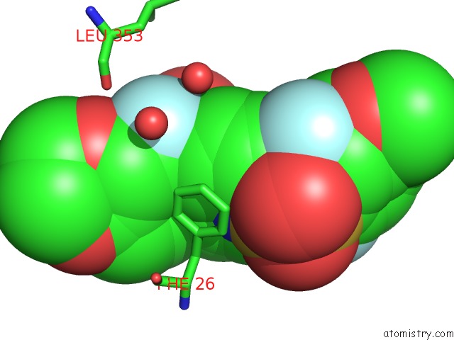
Fluorine binding site 1 out
of 8 in 3gr4
Go back to
Fluorine Binding Sites List in 3gr4
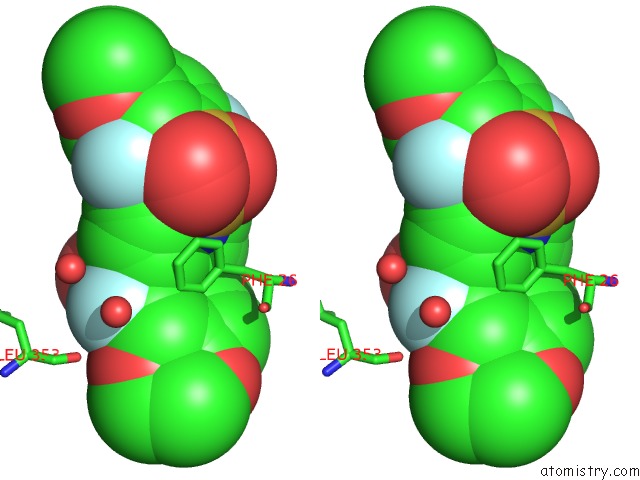
Fluorine binding site 1 out
of 8 in the Activator-Bound Structure of Human Pyruvate Kinase M2
Mono view
Stereo pair view
|
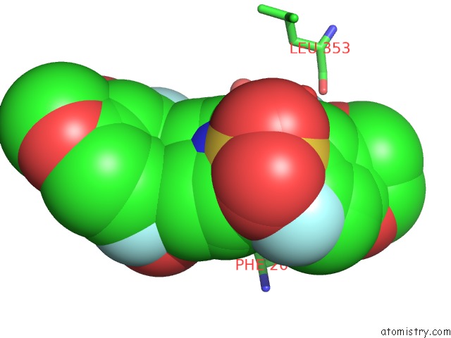
A full contact list of Fluorine with other atoms in the F binding
site number 1 of Activator-Bound Structure of Human Pyruvate Kinase M2 within 5.0Å range:
probe
|
atom
|
residue
|
distance (Å)
|
B
|
Occ
|
A:F550
b:39.9
occ:0.50
|
FAA
|
A:DYY550
|
0.0
|
39.9
|
0.5
|
CAD
|
A:DYY550
|
1.4
|
38.8
|
0.5
|
CAY
|
A:DYY550
|
2.2
|
34.5
|
0.5
|
CAE
|
A:DYY550
|
2.4
|
38.5
|
0.5
|
CAC
|
A:DYY550
|
2.4
|
38.4
|
0.5
|
OAB
|
A:DYY550
|
2.6
|
37.8
|
0.5
|
CAX
|
A:DYY550
|
3.0
|
35.1
|
0.5
|
SAF
|
A:DYY550
|
3.0
|
38.1
|
0.5
|
CAZ
|
A:DYY550
|
3.1
|
33.7
|
0.5
|
OBD
|
A:DYY550
|
3.1
|
36.5
|
0.5
|
CAI
|
A:DYY550
|
3.1
|
38.9
|
0.5
|
SAW
|
A:DYY550
|
3.3
|
36.3
|
0.5
|
OBA
|
A:DYY550
|
3.3
|
34.7
|
0.5
|
O
|
A:LEU353
|
3.5
|
16.9
|
1.0
|
CAH
|
A:DYY550
|
3.5
|
35.9
|
0.5
|
O
|
A:HOH625
|
3.6
|
31.6
|
1.0
|
CE1
|
B:PHE26
|
3.6
|
23.0
|
1.0
|
CAR
|
A:DYY550
|
3.7
|
38.3
|
0.5
|
CAP
|
A:DYY550
|
3.7
|
37.7
|
0.5
|
NAV
|
A:DYY550
|
3.7
|
38.0
|
0.5
|
O
|
A:HOH573
|
3.8
|
22.7
|
1.0
|
NAG
|
A:DYY550
|
3.8
|
36.8
|
0.5
|
CD1
|
B:PHE26
|
4.1
|
23.1
|
1.0
|
CAQ
|
A:DYY550
|
4.2
|
37.9
|
0.5
|
CAK
|
A:DYY550
|
4.2
|
34.1
|
0.5
|
CAM
|
A:DYY550
|
4.2
|
33.4
|
0.5
|
OAS
|
A:DYY550
|
4.3
|
37.9
|
0.5
|
CZ
|
B:PHE26
|
4.4
|
24.2
|
1.0
|
CAH
|
A:DYY550
|
4.5
|
39.9
|
0.5
|
C
|
A:LEU353
|
4.5
|
16.2
|
1.0
|
CAL
|
A:DYY550
|
4.7
|
33.0
|
0.5
|
FBC
|
A:DYY550
|
4.7
|
38.6
|
0.5
|
CBB
|
A:DYY550
|
4.7
|
34.5
|
0.5
|
CAT
|
A:DYY550
|
4.8
|
35.4
|
0.5
|
OAJ
|
A:DYY550
|
4.8
|
35.9
|
0.5
|
CAU
|
A:DYY550
|
4.9
|
38.2
|
0.5
|
|
Fluorine binding site 2 out
of 8 in 3gr4
Go back to
Fluorine Binding Sites List in 3gr4
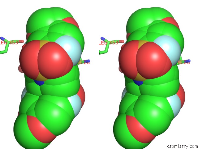
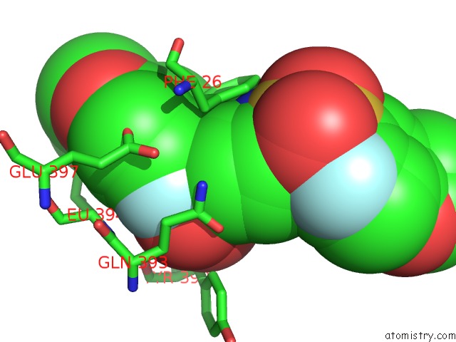
Fluorine binding site 2 out
of 8 in the Activator-Bound Structure of Human Pyruvate Kinase M2
Mono view
Stereo pair view
|
A full contact list of Fluorine with other atoms in the F binding
site number 2 of Activator-Bound Structure of Human Pyruvate Kinase M2 within 5.0Å range:
probe
|
atom
|
residue
|
distance (Å)
|
B
|
Occ
|
A:F550
b:44.6
occ:0.50
|
FAA
|
A:DYY550
|
0.0
|
44.6
|
0.5
|
CAD
|
A:DYY550
|
1.4
|
43.0
|
0.5
|
CAY
|
A:DYY550
|
1.9
|
27.9
|
0.5
|
CAC
|
A:DYY550
|
2.4
|
42.6
|
0.5
|
CAE
|
A:DYY550
|
2.4
|
42.8
|
0.5
|
CAX
|
A:DYY550
|
2.7
|
30.7
|
0.5
|
CAI
|
A:DYY550
|
2.8
|
35.6
|
0.5
|
OBD
|
A:DYY550
|
2.9
|
35.1
|
0.5
|
CAZ
|
A:DYY550
|
2.9
|
26.2
|
0.5
|
OAB
|
A:DYY550
|
3.0
|
43.5
|
0.5
|
SAF
|
A:DYY550
|
3.1
|
43.1
|
0.5
|
SAW
|
A:DYY550
|
3.1
|
34.8
|
0.5
|
OBA
|
A:DYY550
|
3.2
|
26.1
|
0.5
|
CAH
|
A:DYY550
|
3.3
|
39.9
|
0.5
|
NAV
|
A:DYY550
|
3.4
|
35.2
|
0.5
|
CE1
|
A:PHE26
|
3.6
|
23.1
|
1.0
|
O
|
A:HOH695
|
3.6
|
36.4
|
1.0
|
CAR
|
A:DYY550
|
3.6
|
42.0
|
0.5
|
CAP
|
A:DYY550
|
3.7
|
41.9
|
0.5
|
O
|
B:LEU353
|
3.7
|
17.7
|
1.0
|
NAG
|
A:DYY550
|
3.7
|
41.5
|
0.5
|
O
|
B:HOH631
|
3.9
|
32.4
|
1.0
|
CAK
|
A:DYY550
|
4.0
|
27.3
|
0.5
|
CD1
|
A:PHE26
|
4.0
|
22.2
|
1.0
|
CAM
|
A:DYY550
|
4.1
|
25.9
|
0.5
|
CAQ
|
A:DYY550
|
4.1
|
42.0
|
0.5
|
CAH
|
A:DYY550
|
4.2
|
35.9
|
0.5
|
O
|
B:HOH667
|
4.4
|
19.8
|
1.0
|
OAS
|
A:DYY550
|
4.4
|
43.0
|
0.5
|
CAL
|
A:DYY550
|
4.5
|
25.8
|
0.5
|
OAJ
|
A:DYY550
|
4.5
|
34.9
|
0.5
|
CZ
|
A:PHE26
|
4.5
|
23.8
|
1.0
|
CBB
|
A:DYY550
|
4.6
|
26.7
|
0.5
|
CAU
|
A:DYY550
|
4.7
|
35.5
|
0.5
|
C
|
B:LEU353
|
4.7
|
15.7
|
1.0
|
FBC
|
A:DYY550
|
4.7
|
43.0
|
0.5
|
CAT
|
A:DYY550
|
4.7
|
39.8
|
0.5
|
CAI
|
A:DYY550
|
4.8
|
38.9
|
0.5
|
|
Fluorine binding site 3 out
of 8 in 3gr4
Go back to
Fluorine Binding Sites List in 3gr4
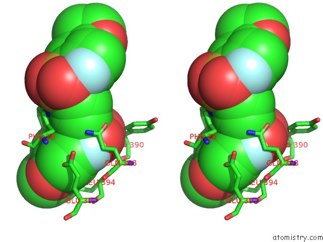
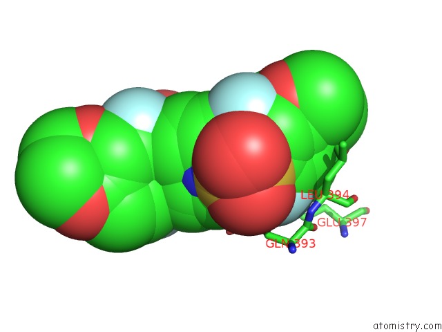
Fluorine binding site 3 out
of 8 in the Activator-Bound Structure of Human Pyruvate Kinase M2
Mono view
Stereo pair view
|
A full contact list of Fluorine with other atoms in the F binding
site number 3 of Activator-Bound Structure of Human Pyruvate Kinase M2 within 5.0Å range:
probe
|
atom
|
residue
|
distance (Å)
|
B
|
Occ
|
A:F550
b:38.6
occ:0.50
|
FBC
|
A:DYY550
|
0.0
|
38.6
|
0.5
|
CAR
|
A:DYY550
|
1.3
|
38.3
|
0.5
|
CAK
|
A:DYY550
|
1.5
|
34.1
|
0.5
|
OAJ
|
A:DYY550
|
1.6
|
35.9
|
0.5
|
CAX
|
A:DYY550
|
1.9
|
35.1
|
0.5
|
SAW
|
A:DYY550
|
2.1
|
36.3
|
0.5
|
CAE
|
A:DYY550
|
2.4
|
38.5
|
0.5
|
CAQ
|
A:DYY550
|
2.4
|
37.9
|
0.5
|
OAS
|
A:DYY550
|
2.6
|
37.9
|
0.5
|
CAL
|
A:DYY550
|
2.8
|
33.0
|
0.5
|
SAF
|
A:DYY550
|
3.0
|
38.1
|
0.5
|
CG
|
A:GLN393
|
3.1
|
20.3
|
1.0
|
CAU
|
A:DYY550
|
3.2
|
38.2
|
0.5
|
NAV
|
A:DYY550
|
3.2
|
38.0
|
0.5
|
CAY
|
A:DYY550
|
3.3
|
34.5
|
0.5
|
CD
|
A:GLN393
|
3.4
|
20.2
|
1.0
|
OBD
|
A:DYY550
|
3.4
|
36.5
|
0.5
|
CAT
|
A:DYY550
|
3.4
|
35.4
|
0.5
|
CB
|
A:GLN393
|
3.5
|
20.4
|
1.0
|
CAP
|
A:DYY550
|
3.6
|
37.7
|
0.5
|
CAD
|
A:DYY550
|
3.6
|
38.8
|
0.5
|
NE2
|
A:GLN393
|
3.6
|
22.2
|
1.0
|
N
|
A:LEU394
|
3.8
|
20.6
|
1.0
|
NAG
|
A:DYY550
|
3.8
|
36.8
|
0.5
|
CAM
|
A:DYY550
|
3.9
|
33.4
|
0.5
|
OE1
|
A:GLN393
|
3.9
|
22.1
|
1.0
|
C
|
A:GLN393
|
4.0
|
18.4
|
1.0
|
CAZ
|
A:DYY550
|
4.1
|
33.7
|
0.5
|
CA
|
A:LEU394
|
4.1
|
20.6
|
1.0
|
CAC
|
A:DYY550
|
4.1
|
38.4
|
0.5
|
OE1
|
A:GLU397
|
4.2
|
24.1
|
1.0
|
OAB
|
A:DYY550
|
4.3
|
37.8
|
0.5
|
CAI
|
A:DYY550
|
4.3
|
38.9
|
0.5
|
O
|
A:GLN393
|
4.3
|
19.9
|
1.0
|
CA
|
A:GLN393
|
4.4
|
19.5
|
1.0
|
CD
|
A:GLU397
|
4.4
|
26.7
|
1.0
|
OE2
|
A:GLU397
|
4.5
|
23.7
|
1.0
|
CB
|
B:PHE26
|
4.5
|
19.0
|
1.0
|
CD1
|
B:PHE26
|
4.5
|
23.1
|
1.0
|
CB
|
A:LEU394
|
4.6
|
19.6
|
1.0
|
O
|
A:TYR390
|
4.6
|
16.7
|
1.0
|
CAT
|
A:DYY550
|
4.7
|
39.8
|
0.5
|
FAA
|
A:DYY550
|
4.7
|
39.9
|
0.5
|
CG
|
B:PHE26
|
4.8
|
22.1
|
1.0
|
CAU
|
A:DYY550
|
4.9
|
35.5
|
0.5
|
CAH
|
A:DYY550
|
4.9
|
35.9
|
0.5
|
CD2
|
A:TYR390
|
4.9
|
17.7
|
1.0
|
|
Fluorine binding site 4 out
of 8 in 3gr4
Go back to
Fluorine Binding Sites List in 3gr4
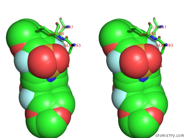
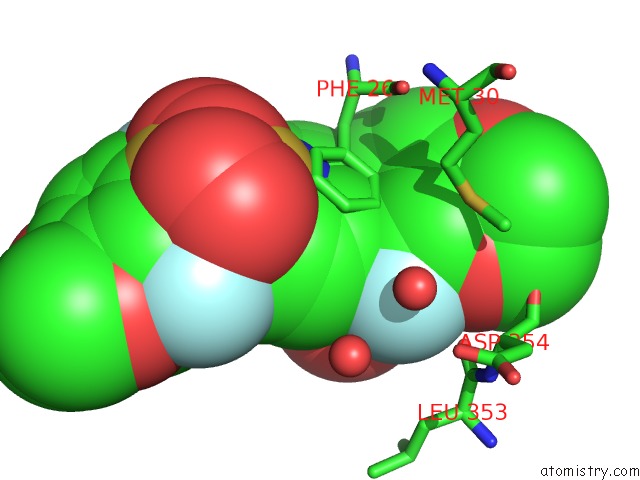
Fluorine binding site 4 out
of 8 in the Activator-Bound Structure of Human Pyruvate Kinase M2
Mono view
Stereo pair view
|
A full contact list of Fluorine with other atoms in the F binding
site number 4 of Activator-Bound Structure of Human Pyruvate Kinase M2 within 5.0Å range:
probe
|
atom
|
residue
|
distance (Å)
|
B
|
Occ
|
A:F550
b:43.0
occ:0.50
|
FBC
|
A:DYY550
|
0.0
|
43.0
|
0.5
|
CAK
|
A:DYY550
|
1.1
|
27.3
|
0.5
|
CAR
|
A:DYY550
|
1.3
|
42.0
|
0.5
|
CAX
|
A:DYY550
|
2.0
|
30.7
|
0.5
|
CAL
|
A:DYY550
|
2.2
|
25.8
|
0.5
|
OAS
|
A:DYY550
|
2.2
|
43.0
|
0.5
|
CAE
|
A:DYY550
|
2.3
|
42.8
|
0.5
|
CAQ
|
A:DYY550
|
2.4
|
42.0
|
0.5
|
OAJ
|
A:DYY550
|
2.5
|
34.9
|
0.5
|
SAW
|
A:DYY550
|
2.7
|
34.8
|
0.5
|
SAF
|
A:DYY550
|
2.9
|
43.1
|
0.5
|
CG
|
B:GLN393
|
3.2
|
21.4
|
1.0
|
CAY
|
A:DYY550
|
3.3
|
27.9
|
0.5
|
CAM
|
A:DYY550
|
3.4
|
25.9
|
0.5
|
CD
|
B:GLN393
|
3.5
|
21.9
|
1.0
|
NE2
|
B:GLN393
|
3.5
|
20.2
|
1.0
|
OE1
|
B:GLU397
|
3.5
|
22.6
|
1.0
|
CAD
|
A:DYY550
|
3.6
|
43.0
|
0.5
|
CAP
|
A:DYY550
|
3.6
|
41.9
|
0.5
|
CB
|
B:GLN393
|
3.7
|
19.6
|
1.0
|
CAU
|
A:DYY550
|
3.7
|
35.5
|
0.5
|
N
|
B:LEU394
|
3.7
|
18.1
|
1.0
|
NAV
|
A:DYY550
|
3.8
|
35.2
|
0.5
|
C
|
B:GLN393
|
3.8
|
18.1
|
1.0
|
CAZ
|
A:DYY550
|
3.8
|
26.2
|
0.5
|
CD
|
B:GLU397
|
3.8
|
23.5
|
1.0
|
CAT
|
A:DYY550
|
3.8
|
39.8
|
0.5
|
CA
|
B:LEU394
|
3.9
|
18.4
|
1.0
|
OBD
|
A:DYY550
|
4.0
|
35.1
|
0.5
|
NAG
|
A:DYY550
|
4.0
|
41.5
|
0.5
|
O
|
B:GLN393
|
4.0
|
18.2
|
1.0
|
OAB
|
A:DYY550
|
4.0
|
43.5
|
0.5
|
CAC
|
A:DYY550
|
4.1
|
42.6
|
0.5
|
OE2
|
B:GLU397
|
4.1
|
21.9
|
1.0
|
CB
|
A:PHE26
|
4.2
|
19.1
|
1.0
|
OE1
|
B:GLN393
|
4.3
|
22.3
|
1.0
|
CA
|
B:GLN393
|
4.4
|
17.7
|
1.0
|
CD1
|
A:PHE26
|
4.4
|
22.2
|
1.0
|
CB
|
B:LEU394
|
4.5
|
18.9
|
1.0
|
CG
|
B:GLU397
|
4.5
|
22.2
|
1.0
|
OAN
|
A:DYY550
|
4.5
|
25.0
|
0.5
|
CAI
|
A:DYY550
|
4.7
|
35.6
|
0.5
|
CG
|
A:PHE26
|
4.7
|
21.7
|
1.0
|
FAA
|
A:DYY550
|
4.7
|
44.6
|
0.5
|
CD1
|
B:LEU394
|
4.9
|
19.7
|
1.0
|
|
Fluorine binding site 5 out
of 8 in 3gr4
Go back to
Fluorine Binding Sites List in 3gr4
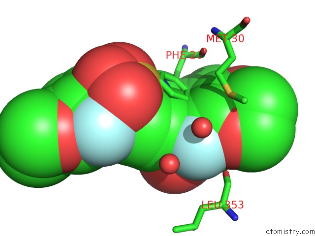
Fluorine binding site 5 out
of 8 in the Activator-Bound Structure of Human Pyruvate Kinase M2
Mono view
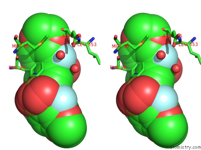
Stereo pair view
|
A full contact list of Fluorine with other atoms in the F binding
site number 5 of Activator-Bound Structure of Human Pyruvate Kinase M2 within 5.0Å range:
probe
|
atom
|
residue
|
distance (Å)
|
B
|
Occ
|
D:F550
b:46.0
occ:0.50
|
FAA
|
D:DYY550
|
0.0
|
46.0
|
0.5
|
CAD
|
D:DYY550
|
1.4
|
45.2
|
0.5
|
CAY
|
D:DYY550
|
2.2
|
32.4
|
0.5
|
CAC
|
D:DYY550
|
2.4
|
44.9
|
0.5
|
CAE
|
D:DYY550
|
2.4
|
44.5
|
0.5
|
OAB
|
D:DYY550
|
2.6
|
43.3
|
0.5
|
CAZ
|
D:DYY550
|
3.0
|
30.7
|
0.5
|
SAF
|
D:DYY550
|
3.0
|
43.5
|
0.5
|
CAI
|
D:DYY550
|
3.1
|
37.0
|
0.5
|
OBA
|
D:DYY550
|
3.1
|
30.8
|
0.5
|
CAX
|
D:DYY550
|
3.2
|
33.8
|
0.5
|
O
|
C:LEU353
|
3.3
|
17.1
|
1.0
|
OBD
|
D:DYY550
|
3.5
|
35.6
|
0.5
|
SAW
|
D:DYY550
|
3.6
|
35.4
|
0.5
|
CE1
|
D:PHE26
|
3.6
|
24.9
|
1.0
|
O
|
C:HOH650
|
3.6
|
30.0
|
1.0
|
O
|
D:HOH655
|
3.7
|
33.0
|
1.0
|
CAP
|
D:DYY550
|
3.7
|
44.8
|
0.5
|
CAH
|
D:DYY550
|
3.7
|
39.7
|
0.5
|
CAR
|
D:DYY550
|
3.7
|
44.5
|
0.5
|
O
|
C:HOH660
|
3.7
|
22.5
|
1.0
|
NAV
|
D:DYY550
|
3.8
|
36.4
|
0.5
|
NAG
|
D:DYY550
|
3.9
|
41.4
|
0.5
|
CD1
|
D:PHE26
|
4.1
|
23.3
|
1.0
|
CAQ
|
D:DYY550
|
4.2
|
44.4
|
0.5
|
CAM
|
D:DYY550
|
4.3
|
31.0
|
0.5
|
OAS
|
D:DYY550
|
4.3
|
42.8
|
0.5
|
C
|
C:LEU353
|
4.3
|
18.2
|
1.0
|
CAK
|
D:DYY550
|
4.4
|
31.0
|
0.5
|
CAH
|
D:DYY550
|
4.4
|
37.7
|
0.5
|
CZ
|
D:PHE26
|
4.5
|
24.4
|
1.0
|
CBB
|
D:DYY550
|
4.5
|
29.8
|
0.5
|
CAL
|
D:DYY550
|
4.8
|
30.5
|
0.5
|
FBC
|
D:DYY550
|
4.8
|
44.3
|
0.5
|
CAT
|
D:DYY550
|
4.9
|
39.7
|
0.5
|
CA
|
C:ASP354
|
4.9
|
15.1
|
1.0
|
SD
|
D:MET30
|
5.0
|
27.1
|
1.0
|
N
|
C:ASP354
|
5.0
|
16.3
|
1.0
|
|
Fluorine binding site 6 out
of 8 in 3gr4
Go back to
Fluorine Binding Sites List in 3gr4
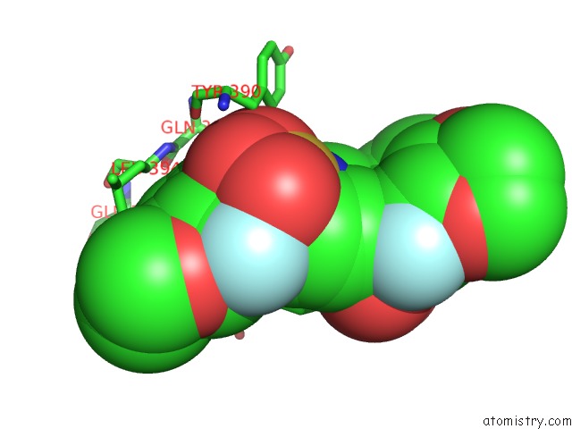
Fluorine binding site 6 out
of 8 in the Activator-Bound Structure of Human Pyruvate Kinase M2
Mono view
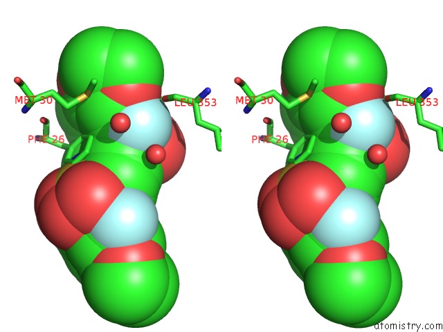
Stereo pair view
|
A full contact list of Fluorine with other atoms in the F binding
site number 6 of Activator-Bound Structure of Human Pyruvate Kinase M2 within 5.0Å range:
probe
|
atom
|
residue
|
distance (Å)
|
B
|
Occ
|
D:F550
b:41.0
occ:0.50
|
FAA
|
D:DYY550
|
0.0
|
41.0
|
0.5
|
CAD
|
D:DYY550
|
1.4
|
40.3
|
0.5
|
CAY
|
D:DYY550
|
1.9
|
32.9
|
0.5
|
CAC
|
D:DYY550
|
2.4
|
39.5
|
0.5
|
CAE
|
D:DYY550
|
2.4
|
40.0
|
0.5
|
CAZ
|
D:DYY550
|
2.8
|
30.8
|
0.5
|
OAB
|
D:DYY550
|
2.8
|
41.1
|
0.5
|
CAX
|
D:DYY550
|
2.9
|
34.1
|
0.5
|
OBA
|
D:DYY550
|
2.9
|
31.2
|
0.5
|
CAI
|
D:DYY550
|
3.0
|
39.1
|
0.5
|
SAF
|
D:DYY550
|
3.1
|
40.7
|
0.5
|
OBD
|
D:DYY550
|
3.3
|
36.9
|
0.5
|
O
|
D:LEU353
|
3.4
|
16.2
|
1.0
|
SAW
|
D:DYY550
|
3.4
|
36.9
|
0.5
|
O
|
D:HOH653
|
3.6
|
32.4
|
1.0
|
CE1
|
C:PHE26
|
3.6
|
23.1
|
1.0
|
CAP
|
D:DYY550
|
3.7
|
39.4
|
0.5
|
CAR
|
D:DYY550
|
3.7
|
38.9
|
0.5
|
CAH
|
D:DYY550
|
3.7
|
37.7
|
0.5
|
NAV
|
D:DYY550
|
3.8
|
38.0
|
0.5
|
O
|
D:HOH595
|
3.9
|
28.0
|
1.0
|
NAG
|
D:DYY550
|
3.9
|
39.0
|
0.5
|
O
|
D:HOH670
|
4.0
|
19.9
|
1.0
|
CD1
|
C:PHE26
|
4.0
|
21.5
|
1.0
|
CAM
|
D:DYY550
|
4.0
|
31.1
|
0.5
|
CAK
|
D:DYY550
|
4.1
|
31.3
|
0.5
|
CAQ
|
D:DYY550
|
4.2
|
38.5
|
0.5
|
CBB
|
D:DYY550
|
4.3
|
30.6
|
0.5
|
CAH
|
D:DYY550
|
4.3
|
39.7
|
0.5
|
C
|
D:LEU353
|
4.4
|
16.6
|
1.0
|
OAS
|
D:DYY550
|
4.4
|
41.2
|
0.5
|
CZ
|
C:PHE26
|
4.5
|
24.0
|
1.0
|
CAL
|
D:DYY550
|
4.6
|
30.3
|
0.5
|
FBC
|
D:DYY550
|
4.8
|
38.9
|
0.5
|
CAT
|
D:DYY550
|
4.8
|
37.7
|
0.5
|
OAJ
|
D:DYY550
|
4.9
|
36.5
|
0.5
|
CAU
|
D:DYY550
|
4.9
|
38.8
|
0.5
|
SD
|
C:MET30
|
4.9
|
25.6
|
1.0
|
|
Fluorine binding site 7 out
of 8 in 3gr4
Go back to
Fluorine Binding Sites List in 3gr4
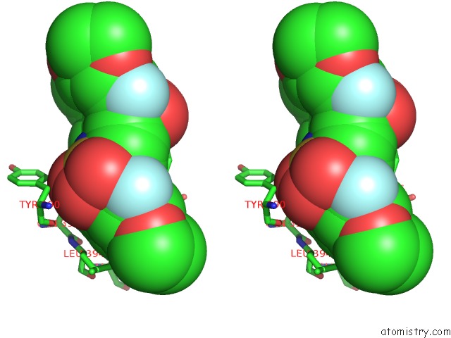
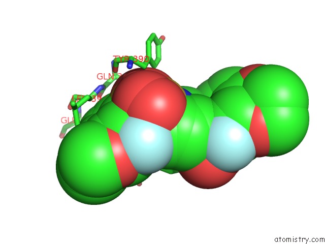
Fluorine binding site 7 out
of 8 in the Activator-Bound Structure of Human Pyruvate Kinase M2
Mono view
Stereo pair view
|
A full contact list of Fluorine with other atoms in the F binding
site number 7 of Activator-Bound Structure of Human Pyruvate Kinase M2 within 5.0Å range:
probe
|
atom
|
residue
|
distance (Å)
|
B
|
Occ
|
D:F550
b:44.3
occ:0.50
|
FBC
|
D:DYY550
|
0.0
|
44.3
|
0.5
|
CAR
|
D:DYY550
|
1.3
|
44.5
|
0.5
|
OAJ
|
D:DYY550
|
1.4
|
35.0
|
0.5
|
CAK
|
D:DYY550
|
1.6
|
31.0
|
0.5
|
CAX
|
D:DYY550
|
1.8
|
33.8
|
0.5
|
SAW
|
D:DYY550
|
1.9
|
35.4
|
0.5
|
CAQ
|
D:DYY550
|
2.4
|
44.4
|
0.5
|
CAE
|
D:DYY550
|
2.4
|
44.5
|
0.5
|
OAS
|
D:DYY550
|
2.7
|
42.8
|
0.5
|
CAL
|
D:DYY550
|
3.0
|
30.5
|
0.5
|
SAF
|
D:DYY550
|
3.0
|
43.5
|
0.5
|
OBD
|
D:DYY550
|
3.1
|
35.6
|
0.5
|
NAV
|
D:DYY550
|
3.1
|
36.4
|
0.5
|
CAU
|
D:DYY550
|
3.1
|
36.8
|
0.5
|
CAY
|
D:DYY550
|
3.2
|
32.4
|
0.5
|
CG
|
C:GLN393
|
3.2
|
18.7
|
1.0
|
CAT
|
D:DYY550
|
3.3
|
39.7
|
0.5
|
CD
|
C:GLN393
|
3.3
|
16.9
|
1.0
|
CB
|
C:GLN393
|
3.5
|
18.4
|
1.0
|
NE2
|
C:GLN393
|
3.6
|
18.9
|
1.0
|
CAP
|
D:DYY550
|
3.6
|
44.8
|
0.5
|
CAD
|
D:DYY550
|
3.7
|
45.2
|
0.5
|
NAG
|
D:DYY550
|
3.7
|
41.4
|
0.5
|
OE1
|
C:GLN393
|
3.9
|
21.2
|
1.0
|
N
|
C:LEU394
|
4.0
|
19.6
|
1.0
|
CAM
|
D:DYY550
|
4.0
|
31.0
|
0.5
|
CAZ
|
D:DYY550
|
4.1
|
30.7
|
0.5
|
C
|
C:GLN393
|
4.1
|
19.0
|
1.0
|
CAI
|
D:DYY550
|
4.1
|
37.0
|
0.5
|
CAC
|
D:DYY550
|
4.1
|
44.9
|
0.5
|
CB
|
D:PHE26
|
4.3
|
20.4
|
1.0
|
CA
|
C:LEU394
|
4.3
|
18.4
|
1.0
|
OE1
|
C:GLU397
|
4.4
|
21.9
|
1.0
|
OAB
|
D:DYY550
|
4.4
|
43.3
|
0.5
|
CA
|
C:GLN393
|
4.4
|
18.4
|
1.0
|
O
|
C:GLN393
|
4.4
|
18.3
|
1.0
|
CD1
|
D:PHE26
|
4.4
|
23.3
|
1.0
|
CD
|
C:GLU397
|
4.5
|
24.1
|
1.0
|
OE2
|
C:GLU397
|
4.6
|
21.9
|
1.0
|
CG
|
D:PHE26
|
4.6
|
22.0
|
1.0
|
CAT
|
D:DYY550
|
4.7
|
37.7
|
0.5
|
O
|
C:TYR390
|
4.7
|
17.4
|
1.0
|
CAU
|
D:DYY550
|
4.7
|
38.8
|
0.5
|
CB
|
C:LEU394
|
4.8
|
18.8
|
1.0
|
FAA
|
D:DYY550
|
4.8
|
46.0
|
0.5
|
CAH
|
D:DYY550
|
4.9
|
39.7
|
0.5
|
CD2
|
C:TYR390
|
5.0
|
20.3
|
1.0
|
|
Fluorine binding site 8 out
of 8 in 3gr4
Go back to
Fluorine Binding Sites List in 3gr4
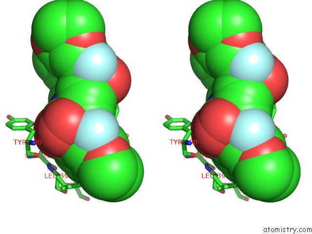
Fluorine binding site 8 out
of 8 in the Activator-Bound Structure of Human Pyruvate Kinase M2
Mono view
Stereo pair view
|
A full contact list of Fluorine with other atoms in the F binding
site number 8 of Activator-Bound Structure of Human Pyruvate Kinase M2 within 5.0Å range:
probe
|
atom
|
residue
|
distance (Å)
|
B
|
Occ
|
D:F550
b:38.9
occ:0.50
|
FBC
|
D:DYY550
|
0.0
|
38.9
|
0.5
|
CAK
|
D:DYY550
|
1.3
|
31.3
|
0.5
|
CAR
|
D:DYY550
|
1.3
|
38.9
|
0.5
|
OAJ
|
D:DYY550
|
1.9
|
36.5
|
0.5
|
CAX
|
D:DYY550
|
1.9
|
34.1
|
0.5
|
SAW
|
D:DYY550
|
2.3
|
36.9
|
0.5
|
CAE
|
D:DYY550
|
2.3
|
40.0
|
0.5
|
CAQ
|
D:DYY550
|
2.4
|
38.5
|
0.5
|
OAS
|
D:DYY550
|
2.4
|
41.2
|
0.5
|
CAL
|
D:DYY550
|
2.6
|
30.3
|
0.5
|
SAF
|
D:DYY550
|
2.9
|
40.7
|
0.5
|
CG
|
D:GLN393
|
3.1
|
17.6
|
1.0
|
CAY
|
D:DYY550
|
3.3
|
32.9
|
0.5
|
CAU
|
D:DYY550
|
3.3
|
38.8
|
0.5
|
NAV
|
D:DYY550
|
3.3
|
38.0
|
0.5
|
CD
|
D:GLN393
|
3.3
|
20.0
|
1.0
|
NE2
|
D:GLN393
|
3.4
|
20.7
|
1.0
|
CAT
|
D:DYY550
|
3.4
|
37.7
|
0.5
|
CB
|
D:GLN393
|
3.5
|
18.1
|
1.0
|
OBD
|
D:DYY550
|
3.5
|
36.9
|
0.5
|
CAP
|
D:DYY550
|
3.6
|
39.4
|
0.5
|
CAD
|
D:DYY550
|
3.6
|
40.3
|
0.5
|
NAG
|
D:DYY550
|
3.7
|
39.0
|
0.5
|
CAM
|
D:DYY550
|
3.7
|
31.1
|
0.5
|
N
|
D:LEU394
|
3.9
|
18.1
|
1.0
|
CAZ
|
D:DYY550
|
4.0
|
30.8
|
0.5
|
OE1
|
D:GLN393
|
4.0
|
23.3
|
1.0
|
C
|
D:GLN393
|
4.0
|
16.8
|
1.0
|
OE1
|
D:GLU397
|
4.1
|
22.4
|
1.0
|
CAC
|
D:DYY550
|
4.1
|
39.5
|
0.5
|
CD
|
D:GLU397
|
4.1
|
23.2
|
1.0
|
OAB
|
D:DYY550
|
4.2
|
41.1
|
0.5
|
CA
|
D:LEU394
|
4.2
|
16.6
|
1.0
|
CB
|
C:PHE26
|
4.2
|
19.0
|
1.0
|
OE2
|
D:GLU397
|
4.3
|
21.2
|
1.0
|
CAI
|
D:DYY550
|
4.3
|
39.1
|
0.5
|
O
|
D:GLN393
|
4.3
|
16.5
|
1.0
|
CD1
|
C:PHE26
|
4.4
|
21.5
|
1.0
|
CA
|
D:GLN393
|
4.4
|
17.8
|
1.0
|
CG
|
C:PHE26
|
4.6
|
21.2
|
1.0
|
CB
|
D:LEU394
|
4.7
|
18.6
|
1.0
|
FAA
|
D:DYY550
|
4.8
|
41.0
|
0.5
|
CAT
|
D:DYY550
|
4.8
|
39.7
|
0.5
|
CAU
|
D:DYY550
|
4.8
|
36.8
|
0.5
|
CG
|
D:GLU397
|
4.9
|
19.8
|
1.0
|
O
|
D:TYR390
|
4.9
|
16.4
|
1.0
|
CAH
|
D:DYY550
|
4.9
|
37.7
|
0.5
|
OAN
|
D:DYY550
|
4.9
|
30.6
|
0.5
|
|
Reference:
B.Hong,
S.Dimov,
W.Tempel,
D.Auld,
C.Thomas,
M.Boxer,
J.-K.Jianq,
A.Skoumbourdis,
S.Min,
N.Southall,
C.H.Arrowsmith,
A.M.Edwards,
C.Bountra,
J.Weigelt,
A.Bochkarev,
J.Inglese,
H.Park.
Activator-Bound Structures of Human Pyruvate Kinase M2 To Be Published.
Page generated: Wed Jul 31 18:58:47 2024
|