Fluorine »
PDB 3gww-3hkw »
3hfg »
Fluorine in PDB 3hfg: Crystal Structure of Human 11-Beta-Hydroxysteroid-Dehydrogenase Bound to An Sulfonyl-Piperazine Inhibitor
Enzymatic activity of Crystal Structure of Human 11-Beta-Hydroxysteroid-Dehydrogenase Bound to An Sulfonyl-Piperazine Inhibitor
All present enzymatic activity of Crystal Structure of Human 11-Beta-Hydroxysteroid-Dehydrogenase Bound to An Sulfonyl-Piperazine Inhibitor:
1.1.1.146;
1.1.1.146;
Protein crystallography data
The structure of Crystal Structure of Human 11-Beta-Hydroxysteroid-Dehydrogenase Bound to An Sulfonyl-Piperazine Inhibitor, PDB code: 3hfg
was solved by
J.Bard,
K.Svenson,
with X-Ray Crystallography technique. A brief refinement statistics is given in the table below:
| Resolution Low / High (Å) | 28.23 / 2.30 |
| Space group | P 1 21 1 |
| Cell size a, b, c (Å), α, β, γ (°) | 56.438, 152.670, 74.203, 90.00, 92.41, 90.00 |
| R / Rfree (%) | 23.1 / 28.5 |
Fluorine Binding Sites:
Pages:
>>> Page 1 <<< Page 2, Binding sites: 11 - 16;Binding sites:
The binding sites of Fluorine atom in the Crystal Structure of Human 11-Beta-Hydroxysteroid-Dehydrogenase Bound to An Sulfonyl-Piperazine Inhibitor (pdb code 3hfg). This binding sites where shown within 5.0 Angstroms radius around Fluorine atom.In total 16 binding sites of Fluorine where determined in the Crystal Structure of Human 11-Beta-Hydroxysteroid-Dehydrogenase Bound to An Sulfonyl-Piperazine Inhibitor, PDB code: 3hfg:
Jump to Fluorine binding site number: 1; 2; 3; 4; 5; 6; 7; 8; 9; 10;
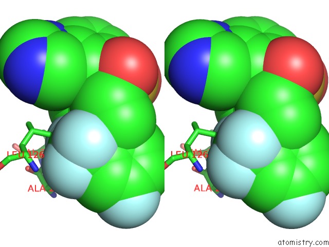
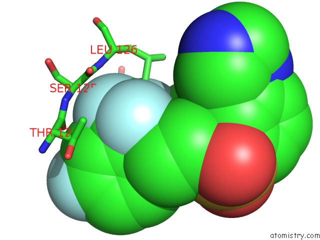
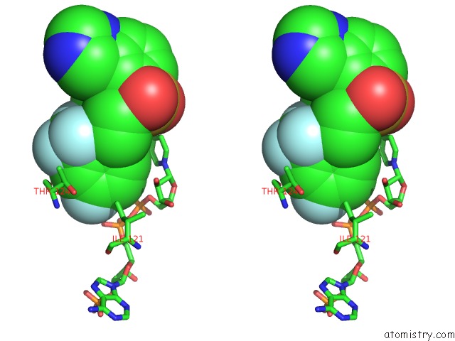
Fluorine binding site 1 out of 16 in 3hfg
Go back to
Fluorine binding site 1 out
of 16 in the Crystal Structure of Human 11-Beta-Hydroxysteroid-Dehydrogenase Bound to An Sulfonyl-Piperazine Inhibitor
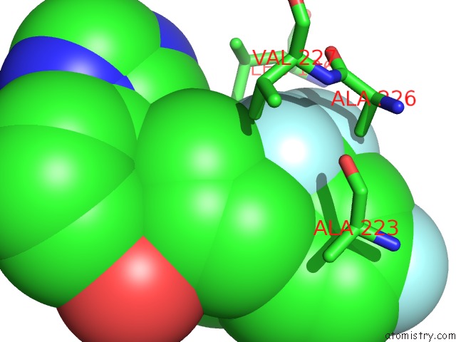
Mono view

Stereo pair view
Mono view
Stereo pair view
A full contact list of Fluorine with other atoms in the F binding
site number 1 of Crystal Structure of Human 11-Beta-Hydroxysteroid-Dehydrogenase Bound to An Sulfonyl-Piperazine Inhibitor within 5.0Å range:
|
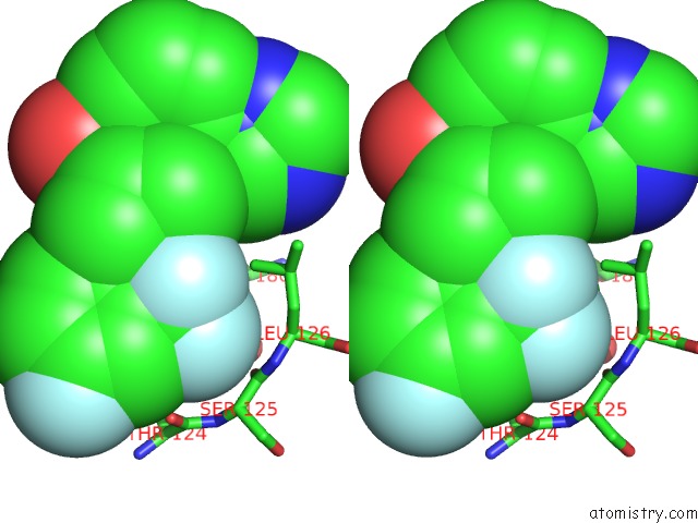
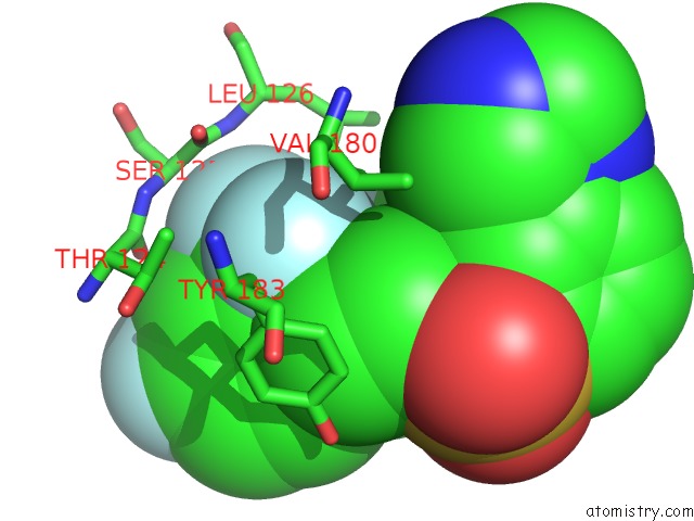
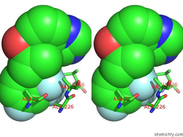
Fluorine binding site 2 out of 16 in 3hfg
Go back to
Fluorine binding site 2 out
of 16 in the Crystal Structure of Human 11-Beta-Hydroxysteroid-Dehydrogenase Bound to An Sulfonyl-Piperazine Inhibitor
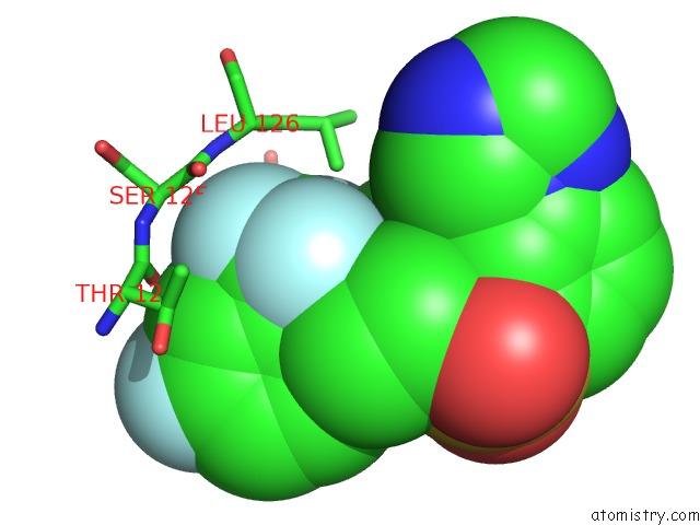
Mono view
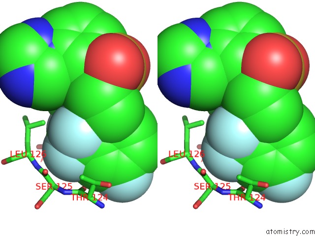
Stereo pair view
Mono view
Stereo pair view
A full contact list of Fluorine with other atoms in the F binding
site number 2 of Crystal Structure of Human 11-Beta-Hydroxysteroid-Dehydrogenase Bound to An Sulfonyl-Piperazine Inhibitor within 5.0Å range:
|
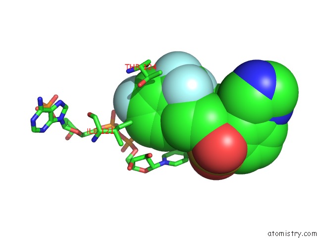
Fluorine binding site 3 out of 16 in 3hfg
Go back to
Fluorine binding site 3 out
of 16 in the Crystal Structure of Human 11-Beta-Hydroxysteroid-Dehydrogenase Bound to An Sulfonyl-Piperazine Inhibitor

Mono view
Stereo pair view
Mono view
Stereo pair view
A full contact list of Fluorine with other atoms in the F binding
site number 3 of Crystal Structure of Human 11-Beta-Hydroxysteroid-Dehydrogenase Bound to An Sulfonyl-Piperazine Inhibitor within 5.0Å range:
|
Fluorine binding site 4 out of 16 in 3hfg
Go back to
Fluorine binding site 4 out
of 16 in the Crystal Structure of Human 11-Beta-Hydroxysteroid-Dehydrogenase Bound to An Sulfonyl-Piperazine Inhibitor

Mono view

Stereo pair view
Mono view
Stereo pair view
A full contact list of Fluorine with other atoms in the F binding
site number 4 of Crystal Structure of Human 11-Beta-Hydroxysteroid-Dehydrogenase Bound to An Sulfonyl-Piperazine Inhibitor within 5.0Å range:
|
Fluorine binding site 5 out of 16 in 3hfg
Go back to
Fluorine binding site 5 out
of 16 in the Crystal Structure of Human 11-Beta-Hydroxysteroid-Dehydrogenase Bound to An Sulfonyl-Piperazine Inhibitor

Mono view
Stereo pair view
Mono view
Stereo pair view
A full contact list of Fluorine with other atoms in the F binding
site number 5 of Crystal Structure of Human 11-Beta-Hydroxysteroid-Dehydrogenase Bound to An Sulfonyl-Piperazine Inhibitor within 5.0Å range:
|
Fluorine binding site 6 out of 16 in 3hfg
Go back to
Fluorine binding site 6 out
of 16 in the Crystal Structure of Human 11-Beta-Hydroxysteroid-Dehydrogenase Bound to An Sulfonyl-Piperazine Inhibitor
Mono view

Stereo pair view
Mono view
Stereo pair view
A full contact list of Fluorine with other atoms in the F binding
site number 6 of Crystal Structure of Human 11-Beta-Hydroxysteroid-Dehydrogenase Bound to An Sulfonyl-Piperazine Inhibitor within 5.0Å range:
|
Fluorine binding site 7 out of 16 in 3hfg
Go back to
Fluorine binding site 7 out
of 16 in the Crystal Structure of Human 11-Beta-Hydroxysteroid-Dehydrogenase Bound to An Sulfonyl-Piperazine Inhibitor
Mono view

Stereo pair view
Mono view
Stereo pair view
A full contact list of Fluorine with other atoms in the F binding
site number 7 of Crystal Structure of Human 11-Beta-Hydroxysteroid-Dehydrogenase Bound to An Sulfonyl-Piperazine Inhibitor within 5.0Å range:
|
Fluorine binding site 8 out of 16 in 3hfg
Go back to
Fluorine binding site 8 out
of 16 in the Crystal Structure of Human 11-Beta-Hydroxysteroid-Dehydrogenase Bound to An Sulfonyl-Piperazine Inhibitor
Mono view
Stereo pair view
Mono view
Stereo pair view
A full contact list of Fluorine with other atoms in the F binding
site number 8 of Crystal Structure of Human 11-Beta-Hydroxysteroid-Dehydrogenase Bound to An Sulfonyl-Piperazine Inhibitor within 5.0Å range:
|
Fluorine binding site 9 out of 16 in 3hfg
Go back to
Fluorine binding site 9 out
of 16 in the Crystal Structure of Human 11-Beta-Hydroxysteroid-Dehydrogenase Bound to An Sulfonyl-Piperazine Inhibitor

Mono view
Stereo pair view
Mono view
Stereo pair view
A full contact list of Fluorine with other atoms in the F binding
site number 9 of Crystal Structure of Human 11-Beta-Hydroxysteroid-Dehydrogenase Bound to An Sulfonyl-Piperazine Inhibitor within 5.0Å range:
|
Fluorine binding site 10 out of 16 in 3hfg
Go back to
Fluorine binding site 10 out
of 16 in the Crystal Structure of Human 11-Beta-Hydroxysteroid-Dehydrogenase Bound to An Sulfonyl-Piperazine Inhibitor

Mono view

Stereo pair view
Mono view
Stereo pair view
A full contact list of Fluorine with other atoms in the F binding
site number 10 of Crystal Structure of Human 11-Beta-Hydroxysteroid-Dehydrogenase Bound to An Sulfonyl-Piperazine Inhibitor within 5.0Å range:
|
Reference:
Z.K.Wan,
E.Chenail,
J.Xiang,
H.Q.Li,
M.Ipek,
J.Bard,
K.Svenson,
T.S.Mansour,
X.Xu,
X.Tian,
V.Suri,
S.Hahm,
Y.Xing,
C.E.Johnson,
X.Li,
A.Qadri,
D.Panza,
M.Perreault,
J.F.Tobin,
E.Saiah.
Efficacious 11BETA-Hydroxysteroid Dehydrogenase Type I Inhibitors in the Diet-Induced Obesity Mouse Model. J.Med.Chem. V. 52 5449 2009.
ISSN: ISSN 0022-2623
PubMed: 19673466
DOI: 10.1021/JM900639U
Page generated: Wed Jul 31 19:13:32 2024
ISSN: ISSN 0022-2623
PubMed: 19673466
DOI: 10.1021/JM900639U
Last articles
Zn in 9J0NZn in 9J0O
Zn in 9J0P
Zn in 9FJX
Zn in 9EKB
Zn in 9C0F
Zn in 9CAH
Zn in 9CH0
Zn in 9CH3
Zn in 9CH1