Fluorine »
PDB 3gww-3hkw »
3hgn »
Fluorine in PDB 3hgn: Structure of Porcine Pancreatic Elastase Complexed with A Potent Peptidyl Inhibitor FR130180 Determined By Neutron Crystallography
Enzymatic activity of Structure of Porcine Pancreatic Elastase Complexed with A Potent Peptidyl Inhibitor FR130180 Determined By Neutron Crystallography
All present enzymatic activity of Structure of Porcine Pancreatic Elastase Complexed with A Potent Peptidyl Inhibitor FR130180 Determined By Neutron Crystallography:
3.4.21.36;
3.4.21.36;
Protein crystallography data
The structure of Structure of Porcine Pancreatic Elastase Complexed with A Potent Peptidyl Inhibitor FR130180 Determined By Neutron Crystallography, PDB code: 3hgn
was solved by
T.Tamada,
T.Kinoshita,
R.Kuroki,
T.Tada,
with X-Ray Crystallography technique. A brief refinement statistics is given in the table below:
| Resolution Low / High (Å) | N/A / 1.65 |
| Space group | P 21 21 21 |
| Cell size a, b, c (Å), α, β, γ (°) | 50.937, 57.464, 75.180, 90.00, 90.00, 90.00 |
| R / Rfree (%) | 14.9 / 16.3 |
Other elements in 3hgn:
The structure of Structure of Porcine Pancreatic Elastase Complexed with A Potent Peptidyl Inhibitor FR130180 Determined By Neutron Crystallography also contains other interesting chemical elements:
| Calcium | (Ca) | 1 atom |
Fluorine Binding Sites:
The binding sites of Fluorine atom in the Structure of Porcine Pancreatic Elastase Complexed with A Potent Peptidyl Inhibitor FR130180 Determined By Neutron Crystallography
(pdb code 3hgn). This binding sites where shown within
5.0 Angstroms radius around Fluorine atom.
In total 3 binding sites of Fluorine where determined in the Structure of Porcine Pancreatic Elastase Complexed with A Potent Peptidyl Inhibitor FR130180 Determined By Neutron Crystallography, PDB code: 3hgn:
Jump to Fluorine binding site number: 1; 2; 3;
In total 3 binding sites of Fluorine where determined in the Structure of Porcine Pancreatic Elastase Complexed with A Potent Peptidyl Inhibitor FR130180 Determined By Neutron Crystallography, PDB code: 3hgn:
Jump to Fluorine binding site number: 1; 2; 3;
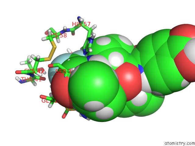
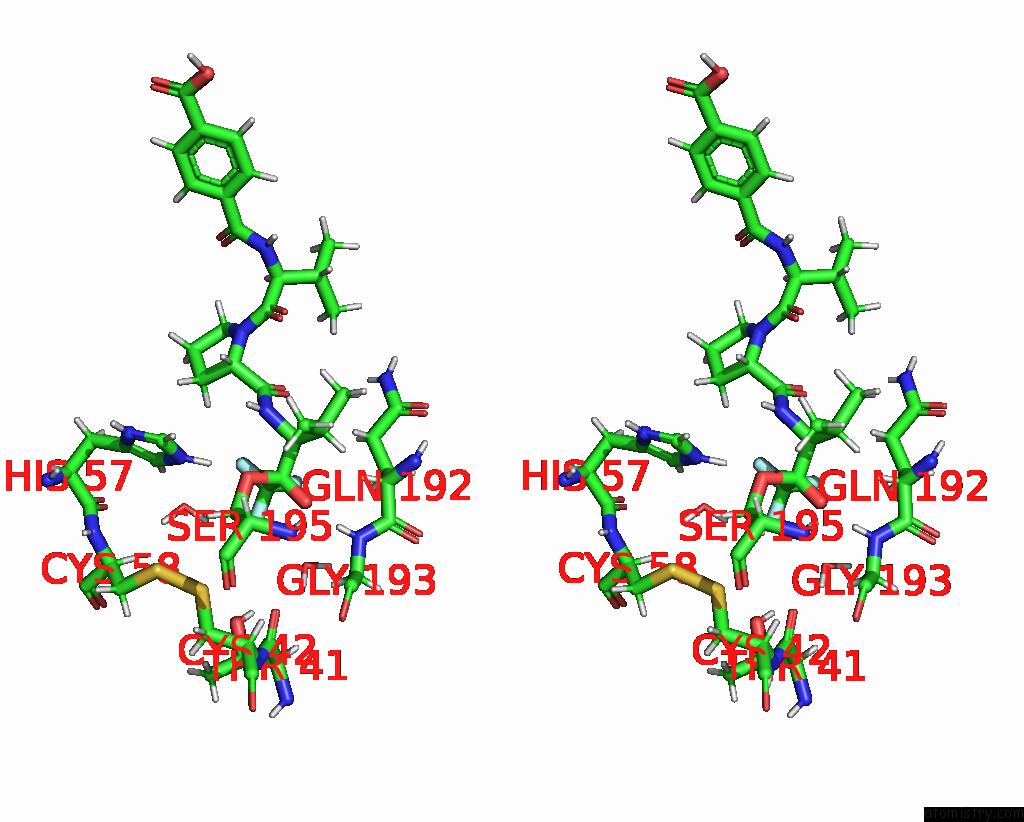
Fluorine binding site 1 out of 3 in 3hgn
Go back to
Fluorine binding site 1 out
of 3 in the Structure of Porcine Pancreatic Elastase Complexed with A Potent Peptidyl Inhibitor FR130180 Determined By Neutron Crystallography
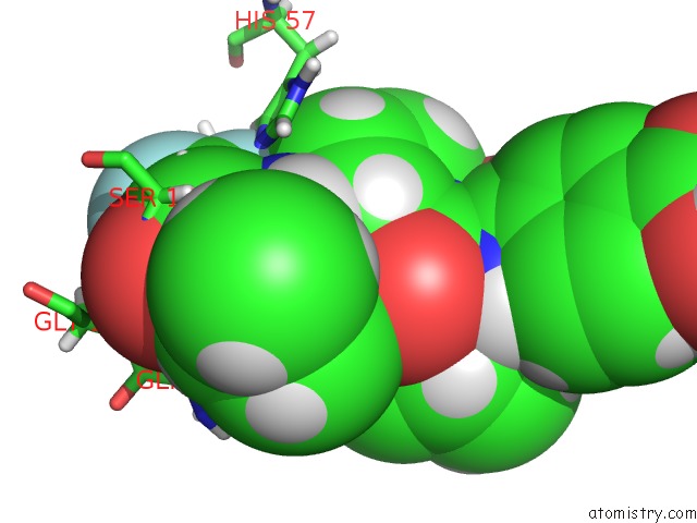
Mono view
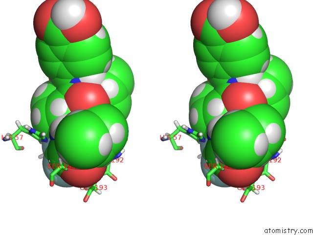
Stereo pair view
Mono view
Stereo pair view
A full contact list of Fluorine with other atoms in the F binding
site number 1 of Structure of Porcine Pancreatic Elastase Complexed with A Potent Peptidyl Inhibitor FR130180 Determined By Neutron Crystallography within 5.0Å range:
|
Fluorine binding site 2 out of 3 in 3hgn
Go back to
Fluorine binding site 2 out
of 3 in the Structure of Porcine Pancreatic Elastase Complexed with A Potent Peptidyl Inhibitor FR130180 Determined By Neutron Crystallography
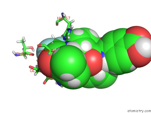
Mono view
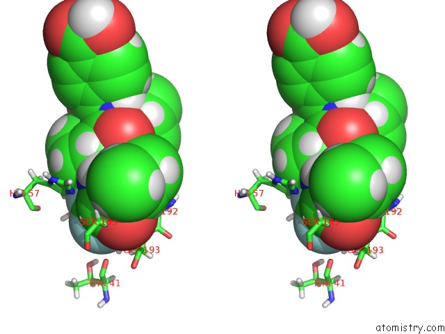
Stereo pair view
Mono view
Stereo pair view
A full contact list of Fluorine with other atoms in the F binding
site number 2 of Structure of Porcine Pancreatic Elastase Complexed with A Potent Peptidyl Inhibitor FR130180 Determined By Neutron Crystallography within 5.0Å range:
|
Fluorine binding site 3 out of 3 in 3hgn
Go back to
Fluorine binding site 3 out
of 3 in the Structure of Porcine Pancreatic Elastase Complexed with A Potent Peptidyl Inhibitor FR130180 Determined By Neutron Crystallography
Mono view
Stereo pair view
Mono view
Stereo pair view
A full contact list of Fluorine with other atoms in the F binding
site number 3 of Structure of Porcine Pancreatic Elastase Complexed with A Potent Peptidyl Inhibitor FR130180 Determined By Neutron Crystallography within 5.0Å range:
|
Reference:
T.Tamada,
T.Kinoshita,
K.Kurihara,
M.Adachi,
T.Ohhara,
K.Imai,
R.Kuroki,
T.Tada.
Combined High-Resolution Neutron and X-Ray Analysis of Inhibited Elastase Confirms the Active-Site Oxyanion Hole But Rules Against A Low-Barrier Hydrogen Bond J.Am.Chem.Soc. V. 131 11033 2009.
ISSN: ISSN 0002-7863
PubMed: 19603802
DOI: 10.1021/JA9028846
Page generated: Wed Jul 31 19:15:23 2024
ISSN: ISSN 0002-7863
PubMed: 19603802
DOI: 10.1021/JA9028846
Last articles
Zn in 9J0NZn in 9J0O
Zn in 9J0P
Zn in 9FJX
Zn in 9EKB
Zn in 9C0F
Zn in 9CAH
Zn in 9CH0
Zn in 9CH3
Zn in 9CH1