Fluorine »
PDB 3hky-3ig6 »
3hvn »
Fluorine in PDB 3hvn: Crystal Structure of Cytotoxin Protein Suilysin From Streptococcus Suis
Protein crystallography data
The structure of Crystal Structure of Cytotoxin Protein Suilysin From Streptococcus Suis, PDB code: 3hvn
was solved by
L.Xu,
B.Huang,
H.Du,
C.X.Zhang,
J.Xu,
X.Li,
Z.Rao,
with X-Ray Crystallography technique. A brief refinement statistics is given in the table below:
| Resolution Low / High (Å) | 28.41 / 2.85 |
| Space group | P 31 2 1 |
| Cell size a, b, c (Å), α, β, γ (°) | 95.720, 95.720, 117.046, 90.00, 90.00, 120.00 |
| R / Rfree (%) | 27.9 / 29.4 |
Fluorine Binding Sites:
The binding sites of Fluorine atom in the Crystal Structure of Cytotoxin Protein Suilysin From Streptococcus Suis
(pdb code 3hvn). This binding sites where shown within
5.0 Angstroms radius around Fluorine atom.
In total 6 binding sites of Fluorine where determined in the Crystal Structure of Cytotoxin Protein Suilysin From Streptococcus Suis, PDB code: 3hvn:
Jump to Fluorine binding site number: 1; 2; 3; 4; 5; 6;
In total 6 binding sites of Fluorine where determined in the Crystal Structure of Cytotoxin Protein Suilysin From Streptococcus Suis, PDB code: 3hvn:
Jump to Fluorine binding site number: 1; 2; 3; 4; 5; 6;
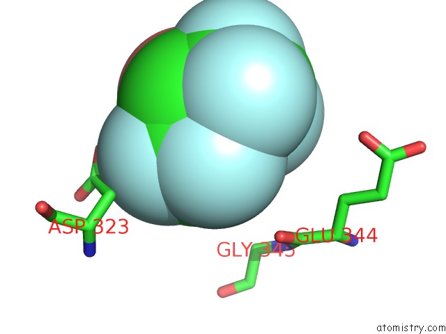
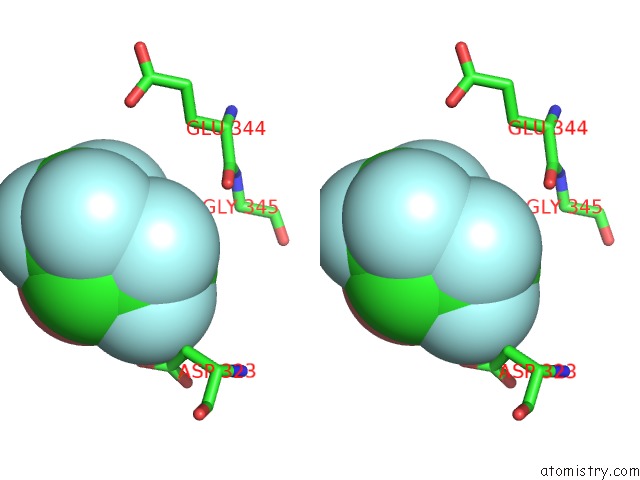
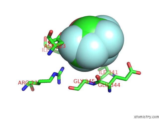
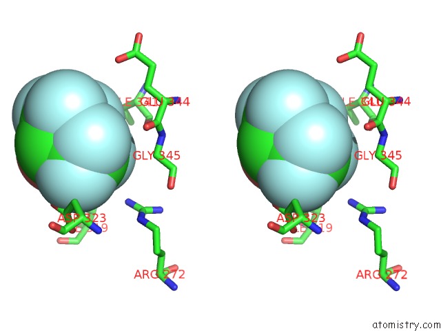
Fluorine binding site 1 out of 6 in 3hvn
Go back to
Fluorine binding site 1 out
of 6 in the Crystal Structure of Cytotoxin Protein Suilysin From Streptococcus Suis
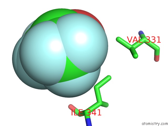
Mono view
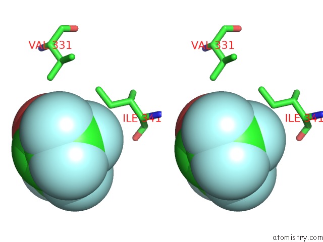
Stereo pair view
Mono view
Stereo pair view
A full contact list of Fluorine with other atoms in the F binding
site number 1 of Crystal Structure of Cytotoxin Protein Suilysin From Streptococcus Suis within 5.0Å range:
|
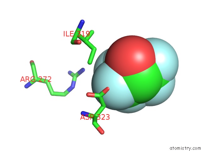
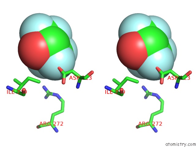
Fluorine binding site 2 out of 6 in 3hvn
Go back to
Fluorine binding site 2 out
of 6 in the Crystal Structure of Cytotoxin Protein Suilysin From Streptococcus Suis
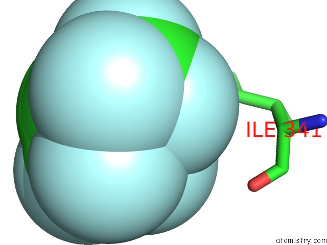
Mono view
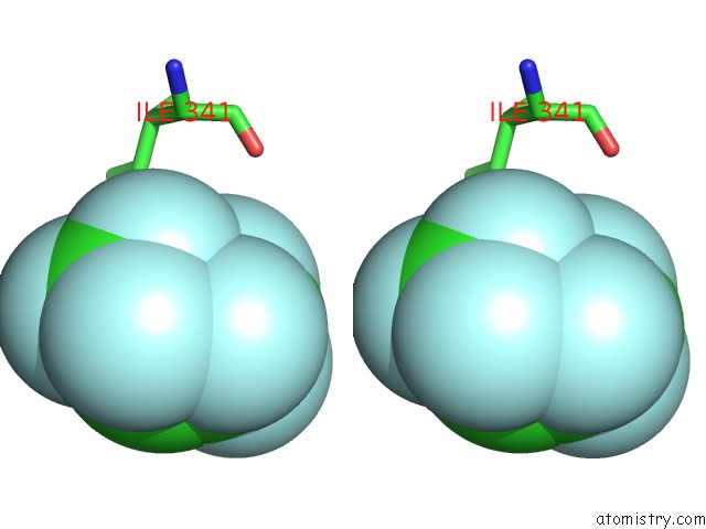
Stereo pair view
Mono view
Stereo pair view
A full contact list of Fluorine with other atoms in the F binding
site number 2 of Crystal Structure of Cytotoxin Protein Suilysin From Streptococcus Suis within 5.0Å range:
|
Fluorine binding site 3 out of 6 in 3hvn
Go back to
Fluorine binding site 3 out
of 6 in the Crystal Structure of Cytotoxin Protein Suilysin From Streptococcus Suis
Mono view
Stereo pair view
Mono view
Stereo pair view
A full contact list of Fluorine with other atoms in the F binding
site number 3 of Crystal Structure of Cytotoxin Protein Suilysin From Streptococcus Suis within 5.0Å range:
|
Fluorine binding site 4 out of 6 in 3hvn
Go back to
Fluorine binding site 4 out
of 6 in the Crystal Structure of Cytotoxin Protein Suilysin From Streptococcus Suis
Mono view
Stereo pair view
Mono view
Stereo pair view
A full contact list of Fluorine with other atoms in the F binding
site number 4 of Crystal Structure of Cytotoxin Protein Suilysin From Streptococcus Suis within 5.0Å range:
|
Fluorine binding site 5 out of 6 in 3hvn
Go back to
Fluorine binding site 5 out
of 6 in the Crystal Structure of Cytotoxin Protein Suilysin From Streptococcus Suis
Mono view
Stereo pair view
Mono view
Stereo pair view
A full contact list of Fluorine with other atoms in the F binding
site number 5 of Crystal Structure of Cytotoxin Protein Suilysin From Streptococcus Suis within 5.0Å range:
|
Fluorine binding site 6 out of 6 in 3hvn
Go back to
Fluorine binding site 6 out
of 6 in the Crystal Structure of Cytotoxin Protein Suilysin From Streptococcus Suis
Mono view
Stereo pair view
Mono view
Stereo pair view
A full contact list of Fluorine with other atoms in the F binding
site number 6 of Crystal Structure of Cytotoxin Protein Suilysin From Streptococcus Suis within 5.0Å range:
|
Reference:
L.Xu,
B.Huang,
H.Du,
X.C.Zhang,
J.Xu,
X.Li,
Z.Rao.
Crystal Structure of Cytotoxin Protein Suilysin From Streptococcus Suis. Protein Cell V. 1 96 2010.
ISSN: ESSN 1674-8018
PubMed: 21204001
DOI: 10.1007/S13238-010-0012-3
Page generated: Wed Jul 31 19:24:19 2024
ISSN: ESSN 1674-8018
PubMed: 21204001
DOI: 10.1007/S13238-010-0012-3
Last articles
Zn in 9MJ5Zn in 9HNW
Zn in 9G0L
Zn in 9FNE
Zn in 9DZN
Zn in 9E0I
Zn in 9D32
Zn in 9DAK
Zn in 8ZXC
Zn in 8ZUF