Fluorine in PDB 3k5v: Structure of Abl Kinase in Complex with Imatinib and Gnf-2
Enzymatic activity of Structure of Abl Kinase in Complex with Imatinib and Gnf-2
All present enzymatic activity of Structure of Abl Kinase in Complex with Imatinib and Gnf-2:
2.7.10.2;
Protein crystallography data
The structure of Structure of Abl Kinase in Complex with Imatinib and Gnf-2, PDB code: 3k5v
was solved by
S.W.Cowan-Jacob,
G.Fendrich,
G.Rummel,
A.Strauss,
with X-Ray Crystallography technique. A brief refinement statistics is given in the table below:
Resolution Low / High (Å)
|
38.95 /
1.74
|
Space group
|
P 1
|
Cell size a, b, c (Å), α, β, γ (°)
|
42.070,
65.268,
66.259,
72.82,
80.25,
84.86
|
R / Rfree (%)
|
19.9 /
23.1
|
Other elements in 3k5v:
The structure of Structure of Abl Kinase in Complex with Imatinib and Gnf-2 also contains other interesting chemical elements:
Fluorine Binding Sites:
The binding sites of Fluorine atom in the Structure of Abl Kinase in Complex with Imatinib and Gnf-2
(pdb code 3k5v). This binding sites where shown within
5.0 Angstroms radius around Fluorine atom.
In total 6 binding sites of Fluorine where determined in the
Structure of Abl Kinase in Complex with Imatinib and Gnf-2, PDB code: 3k5v:
Jump to Fluorine binding site number:
1;
2;
3;
4;
5;
6;
Fluorine binding site 1 out
of 6 in 3k5v
Go back to
Fluorine Binding Sites List in 3k5v
Fluorine binding site 1 out
of 6 in the Structure of Abl Kinase in Complex with Imatinib and Gnf-2
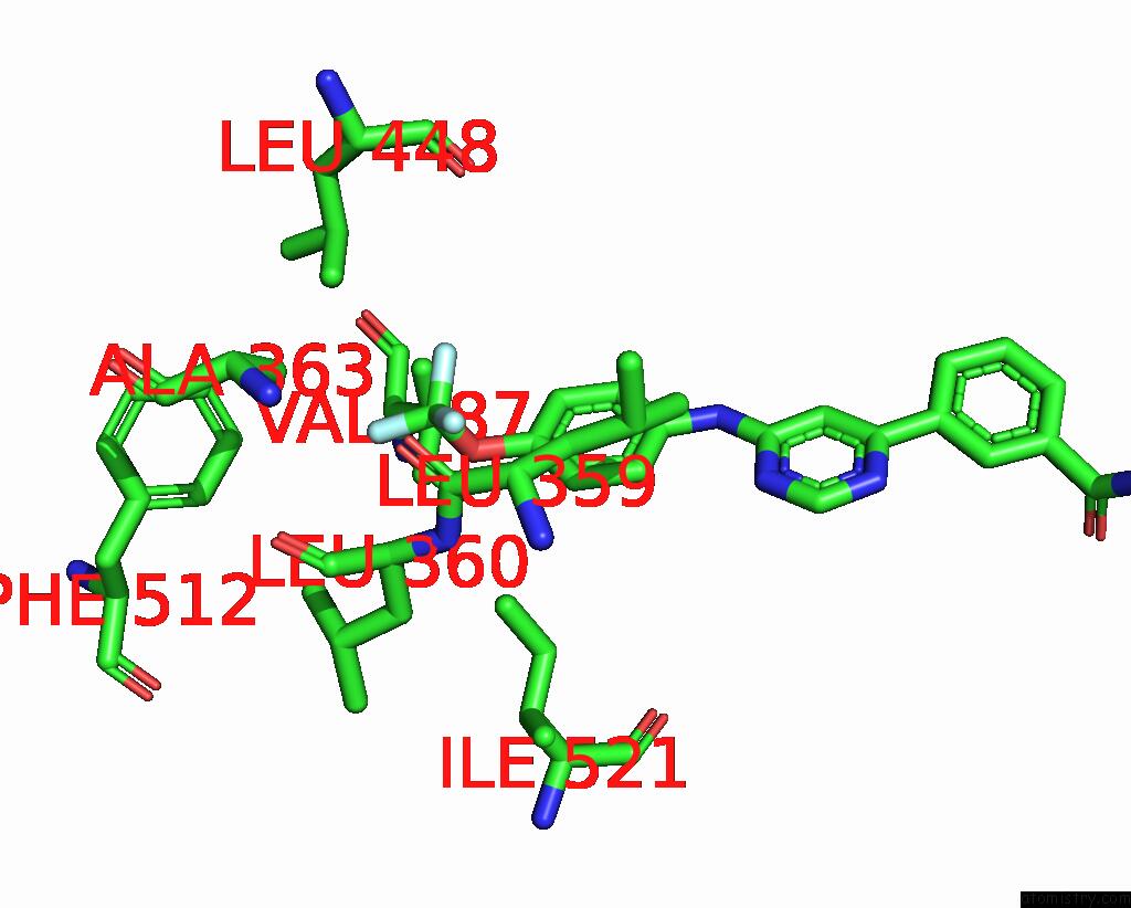
Mono view
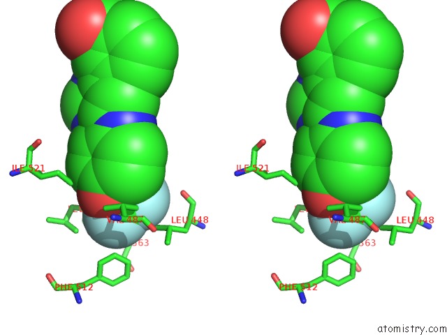
Stereo pair view
|
A full contact list of Fluorine with other atoms in the F binding
site number 1 of Structure of Abl Kinase in Complex with Imatinib and Gnf-2 within 5.0Å range:
probe
|
atom
|
residue
|
distance (Å)
|
B
|
Occ
|
A:F1
b:26.0
occ:1.00
|
F1
|
A:STJ1
|
0.0
|
26.0
|
1.0
|
C2
|
A:STJ1
|
1.4
|
25.0
|
1.0
|
O5
|
A:STJ1
|
2.2
|
24.2
|
1.0
|
F4
|
A:STJ1
|
2.2
|
25.3
|
1.0
|
F3
|
A:STJ1
|
2.2
|
26.0
|
1.0
|
CB
|
A:ALA363
|
3.4
|
14.7
|
1.0
|
C6
|
A:STJ1
|
3.5
|
23.3
|
1.0
|
CE1
|
A:PHE512
|
3.6
|
18.7
|
1.0
|
CD1
|
A:LEU448
|
3.7
|
17.9
|
1.0
|
CA
|
A:LEU360
|
3.8
|
15.8
|
1.0
|
CD1
|
A:LEU360
|
3.8
|
18.6
|
1.0
|
CG1
|
A:VAL487
|
4.2
|
17.2
|
1.0
|
CZ
|
A:PHE512
|
4.2
|
16.8
|
1.0
|
O
|
A:LEU359
|
4.3
|
15.4
|
1.0
|
CB
|
A:LEU360
|
4.3
|
16.4
|
1.0
|
N
|
A:LEU360
|
4.3
|
15.9
|
1.0
|
C14
|
A:STJ1
|
4.4
|
25.6
|
1.0
|
CG2
|
A:VAL487
|
4.4
|
19.5
|
1.0
|
C7
|
A:STJ1
|
4.4
|
24.9
|
1.0
|
CD1
|
A:PHE512
|
4.4
|
21.1
|
1.0
|
O
|
A:LEU360
|
4.6
|
15.0
|
1.0
|
C
|
A:LEU359
|
4.6
|
16.0
|
1.0
|
C
|
A:LEU360
|
4.7
|
15.3
|
1.0
|
CG
|
A:LEU360
|
4.7
|
17.8
|
1.0
|
CG
|
A:LEU448
|
4.9
|
17.8
|
1.0
|
CA
|
A:ALA363
|
4.9
|
14.1
|
1.0
|
CD1
|
A:ILE521
|
4.9
|
23.2
|
1.0
|
CB
|
A:VAL487
|
5.0
|
17.9
|
1.0
|
|
Fluorine binding site 2 out
of 6 in 3k5v
Go back to
Fluorine Binding Sites List in 3k5v
Fluorine binding site 2 out
of 6 in the Structure of Abl Kinase in Complex with Imatinib and Gnf-2
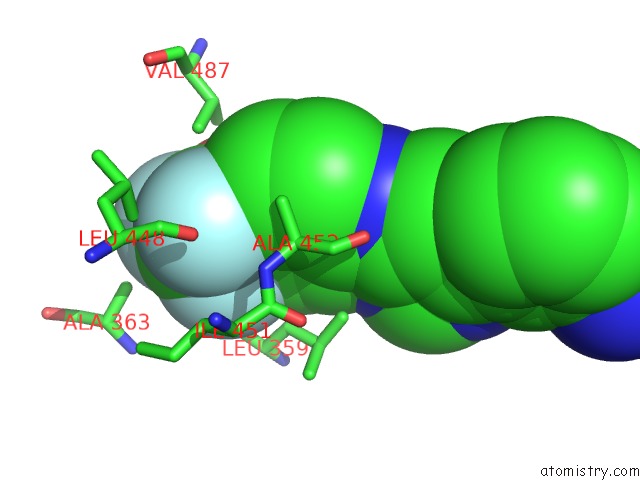
Mono view
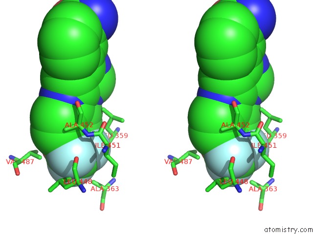
Stereo pair view
|
A full contact list of Fluorine with other atoms in the F binding
site number 2 of Structure of Abl Kinase in Complex with Imatinib and Gnf-2 within 5.0Å range:
probe
|
atom
|
residue
|
distance (Å)
|
B
|
Occ
|
A:F1
b:26.0
occ:1.00
|
F3
|
A:STJ1
|
0.0
|
26.0
|
1.0
|
C2
|
A:STJ1
|
1.4
|
25.0
|
1.0
|
F4
|
A:STJ1
|
2.2
|
25.3
|
1.0
|
F1
|
A:STJ1
|
2.2
|
26.0
|
1.0
|
O5
|
A:STJ1
|
2.3
|
24.2
|
1.0
|
C6
|
A:STJ1
|
2.9
|
23.3
|
1.0
|
C14
|
A:STJ1
|
3.1
|
25.6
|
1.0
|
CD1
|
A:LEU448
|
3.1
|
17.9
|
1.0
|
CG1
|
A:VAL487
|
3.7
|
17.2
|
1.0
|
CG2
|
A:ILE451
|
4.0
|
14.6
|
1.0
|
C7
|
A:STJ1
|
4.0
|
24.9
|
1.0
|
CG
|
A:LEU448
|
4.0
|
17.8
|
1.0
|
O
|
A:LEU448
|
4.1
|
14.0
|
1.0
|
C12
|
A:STJ1
|
4.3
|
25.8
|
1.0
|
CB
|
A:ALA363
|
4.4
|
14.7
|
1.0
|
CB
|
A:ALA452
|
4.5
|
16.1
|
1.0
|
N
|
A:ALA452
|
4.5
|
14.8
|
1.0
|
CB
|
A:ILE451
|
4.5
|
14.4
|
1.0
|
CA
|
A:ALA452
|
4.7
|
16.0
|
1.0
|
O
|
A:LEU359
|
4.8
|
15.4
|
1.0
|
CG2
|
A:VAL487
|
4.9
|
19.5
|
1.0
|
CB
|
A:VAL487
|
4.9
|
17.9
|
1.0
|
C
|
A:LEU448
|
4.9
|
14.3
|
1.0
|
C9
|
A:STJ1
|
5.0
|
25.0
|
1.0
|
|
Fluorine binding site 3 out
of 6 in 3k5v
Go back to
Fluorine Binding Sites List in 3k5v
Fluorine binding site 3 out
of 6 in the Structure of Abl Kinase in Complex with Imatinib and Gnf-2
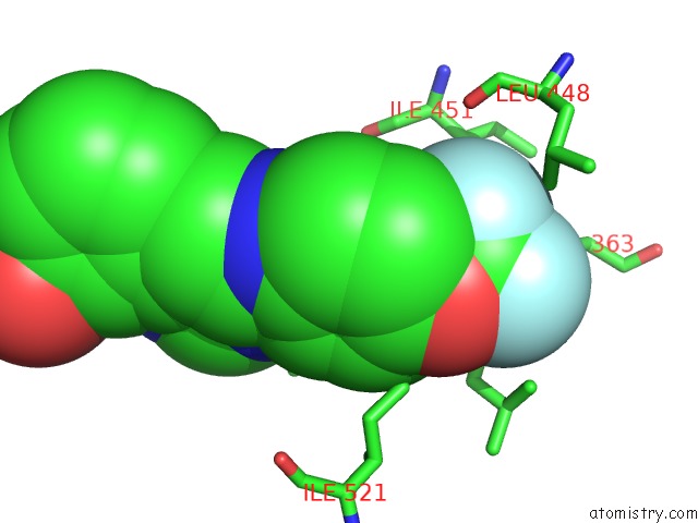
Mono view
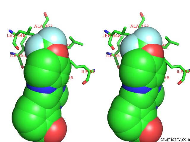
Stereo pair view
|
A full contact list of Fluorine with other atoms in the F binding
site number 3 of Structure of Abl Kinase in Complex with Imatinib and Gnf-2 within 5.0Å range:
probe
|
atom
|
residue
|
distance (Å)
|
B
|
Occ
|
A:F1
b:25.3
occ:1.00
|
F4
|
A:STJ1
|
0.0
|
25.3
|
1.0
|
C2
|
A:STJ1
|
1.4
|
25.0
|
1.0
|
F1
|
A:STJ1
|
2.2
|
26.0
|
1.0
|
F3
|
A:STJ1
|
2.2
|
26.0
|
1.0
|
O5
|
A:STJ1
|
2.3
|
24.2
|
1.0
|
C6
|
A:STJ1
|
2.9
|
23.3
|
1.0
|
O
|
A:LEU359
|
2.9
|
15.4
|
1.0
|
C
|
A:LEU359
|
2.9
|
16.0
|
1.0
|
N
|
A:LEU360
|
3.1
|
15.9
|
1.0
|
C7
|
A:STJ1
|
3.2
|
24.9
|
1.0
|
CA
|
A:LEU360
|
3.3
|
15.8
|
1.0
|
CB
|
A:LEU359
|
3.5
|
16.3
|
1.0
|
CG2
|
A:ILE451
|
3.5
|
14.6
|
1.0
|
CA
|
A:LEU359
|
3.8
|
15.7
|
1.0
|
C14
|
A:STJ1
|
3.9
|
25.6
|
1.0
|
CB
|
A:ALA363
|
4.0
|
14.7
|
1.0
|
CB
|
A:LEU360
|
4.2
|
16.4
|
1.0
|
C9
|
A:STJ1
|
4.4
|
25.0
|
1.0
|
C
|
A:LEU360
|
4.5
|
15.3
|
1.0
|
CD1
|
A:LEU448
|
4.7
|
17.9
|
1.0
|
O
|
A:ALA356
|
4.7
|
18.4
|
1.0
|
CB
|
A:ILE451
|
4.7
|
14.4
|
1.0
|
O
|
A:LEU360
|
4.8
|
15.0
|
1.0
|
CG
|
A:LEU359
|
4.8
|
17.7
|
1.0
|
C12
|
A:STJ1
|
4.9
|
25.8
|
1.0
|
CD1
|
A:LEU360
|
4.9
|
18.6
|
1.0
|
CD1
|
A:ILE521
|
5.0
|
23.2
|
1.0
|
N
|
A:LEU359
|
5.0
|
15.0
|
1.0
|
|
Fluorine binding site 4 out
of 6 in 3k5v
Go back to
Fluorine Binding Sites List in 3k5v
Fluorine binding site 4 out
of 6 in the Structure of Abl Kinase in Complex with Imatinib and Gnf-2
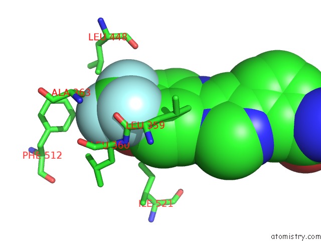
Mono view
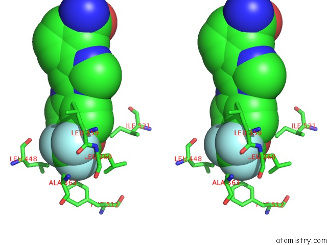
Stereo pair view
|
A full contact list of Fluorine with other atoms in the F binding
site number 4 of Structure of Abl Kinase in Complex with Imatinib and Gnf-2 within 5.0Å range:
probe
|
atom
|
residue
|
distance (Å)
|
B
|
Occ
|
B:F1
b:50.9
occ:1.00
|
F1
|
B:STJ1
|
0.0
|
50.9
|
1.0
|
C2
|
B:STJ1
|
1.4
|
51.9
|
1.0
|
F3
|
B:STJ1
|
2.2
|
51.3
|
1.0
|
F4
|
B:STJ1
|
2.2
|
49.1
|
1.0
|
O5
|
B:STJ1
|
2.3
|
53.1
|
1.0
|
CE1
|
B:PHE512
|
3.6
|
43.3
|
1.0
|
C6
|
B:STJ1
|
3.6
|
55.3
|
1.0
|
CB
|
B:ALA363
|
3.7
|
29.5
|
1.0
|
CA
|
B:LEU360
|
3.7
|
34.2
|
1.0
|
CD1
|
B:LEU448
|
3.8
|
29.7
|
1.0
|
CD1
|
B:LEU360
|
3.9
|
34.4
|
1.0
|
CG1
|
B:VAL487
|
4.2
|
33.1
|
1.0
|
CB
|
B:LEU360
|
4.2
|
34.6
|
1.0
|
CZ
|
B:PHE512
|
4.2
|
42.6
|
1.0
|
N
|
B:LEU360
|
4.3
|
34.5
|
1.0
|
CG2
|
B:VAL487
|
4.3
|
34.6
|
1.0
|
CD1
|
B:PHE512
|
4.3
|
44.0
|
1.0
|
O
|
B:LEU359
|
4.3
|
34.6
|
1.0
|
C14
|
B:STJ1
|
4.4
|
56.3
|
1.0
|
C7
|
B:STJ1
|
4.5
|
56.3
|
1.0
|
C
|
B:LEU359
|
4.5
|
35.1
|
1.0
|
O
|
B:LEU360
|
4.7
|
33.6
|
1.0
|
C
|
B:LEU360
|
4.7
|
33.8
|
1.0
|
CG
|
B:LEU360
|
4.7
|
35.0
|
1.0
|
CB
|
B:VAL487
|
4.9
|
33.3
|
1.0
|
CG
|
B:LEU448
|
4.9
|
28.4
|
1.0
|
CD1
|
B:ILE521
|
5.0
|
59.9
|
1.0
|
|
Fluorine binding site 5 out
of 6 in 3k5v
Go back to
Fluorine Binding Sites List in 3k5v
Fluorine binding site 5 out
of 6 in the Structure of Abl Kinase in Complex with Imatinib and Gnf-2
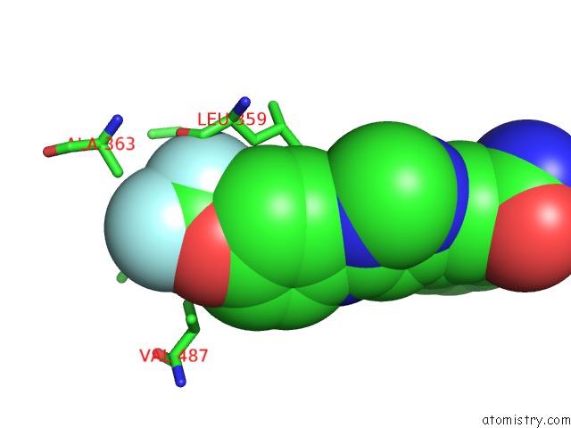
Mono view
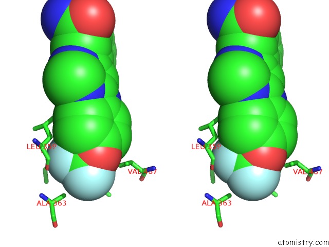
Stereo pair view
|
A full contact list of Fluorine with other atoms in the F binding
site number 5 of Structure of Abl Kinase in Complex with Imatinib and Gnf-2 within 5.0Å range:
probe
|
atom
|
residue
|
distance (Å)
|
B
|
Occ
|
B:F1
b:51.3
occ:1.00
|
F3
|
B:STJ1
|
0.0
|
51.3
|
1.0
|
C2
|
B:STJ1
|
1.4
|
51.9
|
1.0
|
F4
|
B:STJ1
|
2.2
|
49.1
|
1.0
|
F1
|
B:STJ1
|
2.2
|
50.9
|
1.0
|
O5
|
B:STJ1
|
2.3
|
53.1
|
1.0
|
C6
|
B:STJ1
|
2.9
|
55.3
|
1.0
|
C14
|
B:STJ1
|
3.2
|
56.3
|
1.0
|
CD1
|
B:LEU448
|
3.2
|
29.7
|
1.0
|
CG1
|
B:VAL487
|
3.6
|
33.1
|
1.0
|
CG
|
B:LEU448
|
4.0
|
28.4
|
1.0
|
C7
|
B:STJ1
|
4.0
|
56.3
|
1.0
|
CG2
|
B:ILE451
|
4.0
|
28.5
|
1.0
|
O
|
B:LEU448
|
4.1
|
26.9
|
1.0
|
CB
|
B:ALA452
|
4.4
|
30.1
|
1.0
|
C12
|
B:STJ1
|
4.4
|
57.1
|
1.0
|
N
|
B:ALA452
|
4.5
|
30.1
|
1.0
|
CB
|
B:ALA363
|
4.5
|
29.5
|
1.0
|
CB
|
B:ILE451
|
4.6
|
28.1
|
1.0
|
CA
|
B:ALA452
|
4.6
|
31.1
|
1.0
|
CG2
|
B:VAL487
|
4.7
|
34.6
|
1.0
|
CB
|
B:VAL487
|
4.7
|
33.3
|
1.0
|
O
|
B:LEU359
|
4.9
|
34.6
|
1.0
|
C
|
B:LEU448
|
4.9
|
26.6
|
1.0
|
CD2
|
B:LEU448
|
5.0
|
27.9
|
1.0
|
|
Fluorine binding site 6 out
of 6 in 3k5v
Go back to
Fluorine Binding Sites List in 3k5v
Fluorine binding site 6 out
of 6 in the Structure of Abl Kinase in Complex with Imatinib and Gnf-2
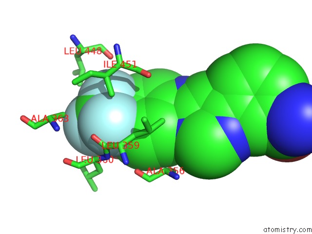
Mono view
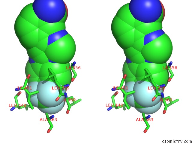
Stereo pair view
|
A full contact list of Fluorine with other atoms in the F binding
site number 6 of Structure of Abl Kinase in Complex with Imatinib and Gnf-2 within 5.0Å range:
probe
|
atom
|
residue
|
distance (Å)
|
B
|
Occ
|
B:F1
b:49.1
occ:1.00
|
F4
|
B:STJ1
|
0.0
|
49.1
|
1.0
|
C2
|
B:STJ1
|
1.4
|
51.9
|
1.0
|
F3
|
B:STJ1
|
2.2
|
51.3
|
1.0
|
F1
|
B:STJ1
|
2.2
|
50.9
|
1.0
|
O5
|
B:STJ1
|
2.3
|
53.1
|
1.0
|
C6
|
B:STJ1
|
2.9
|
55.3
|
1.0
|
C
|
B:LEU359
|
3.0
|
35.1
|
1.0
|
O
|
B:LEU359
|
3.0
|
34.6
|
1.0
|
N
|
B:LEU360
|
3.1
|
34.5
|
1.0
|
C7
|
B:STJ1
|
3.3
|
56.3
|
1.0
|
CB
|
B:LEU359
|
3.4
|
35.4
|
1.0
|
CA
|
B:LEU360
|
3.4
|
34.2
|
1.0
|
CG2
|
B:ILE451
|
3.5
|
28.5
|
1.0
|
CA
|
B:LEU359
|
3.8
|
35.3
|
1.0
|
C14
|
B:STJ1
|
3.9
|
56.3
|
1.0
|
CB
|
B:ALA363
|
4.2
|
29.5
|
1.0
|
CB
|
B:LEU360
|
4.3
|
34.6
|
1.0
|
C9
|
B:STJ1
|
4.4
|
57.2
|
1.0
|
C
|
B:LEU360
|
4.6
|
33.8
|
1.0
|
CB
|
B:ILE451
|
4.7
|
28.1
|
1.0
|
CG
|
B:LEU359
|
4.7
|
35.2
|
1.0
|
O
|
B:ALA356
|
4.7
|
43.2
|
1.0
|
CD1
|
B:LEU448
|
4.7
|
29.7
|
1.0
|
CD1
|
B:LEU359
|
4.9
|
35.4
|
1.0
|
C12
|
B:STJ1
|
4.9
|
57.1
|
1.0
|
N
|
B:LEU359
|
5.0
|
36.4
|
1.0
|
|
Reference:
J.Zhang,
F.J.Adrian,
W.Jahnke,
S.W.Cowan-Jacob,
A.G.Li,
R.E.Iacob,
T.Sim,
J.Powers,
C.Dierks,
F.Sun,
G.R.Guo,
Q.Ding,
B.Okram,
Y.Choi,
A.Wojciechowski,
X.Deng,
G.Liu,
G.Fendrich,
A.Strauss,
N.Vajpai,
S.Grzesiek,
T.Tuntland,
Y.Liu,
B.Bursulaya,
M.Azam,
P.W.Manley,
J.R.Engen,
G.Q.Daley,
M.Warmuth,
N.S.Gray.
Targeting Bcr-Abl By Combining Allosteric with Atp-Binding-Site Inhibitors. Nature V. 463 501 2010.
ISSN: ISSN 0028-0836
PubMed: 20072125
DOI: 10.1038/NATURE08675
Page generated: Wed Jul 31 19:47:29 2024
|