Fluorine »
PDB 3jx3-3kql »
3kk6 »
Fluorine in PDB 3kk6: Crystal Structure of Cyclooxygenase-1 in Complex with Celecoxib
Enzymatic activity of Crystal Structure of Cyclooxygenase-1 in Complex with Celecoxib
All present enzymatic activity of Crystal Structure of Cyclooxygenase-1 in Complex with Celecoxib:
1.14.99.1;
1.14.99.1;
Protein crystallography data
The structure of Crystal Structure of Cyclooxygenase-1 in Complex with Celecoxib, PDB code: 3kk6
was solved by
R.S.Sidhu,
with X-Ray Crystallography technique. A brief refinement statistics is given in the table below:
| Resolution Low / High (Å) | 41.42 / 2.75 |
| Space group | P 65 |
| Cell size a, b, c (Å), α, β, γ (°) | 181.035, 181.035, 102.698, 90.00, 90.00, 120.00 |
| R / Rfree (%) | 20.7 / 24.2 |
Other elements in 3kk6:
The structure of Crystal Structure of Cyclooxygenase-1 in Complex with Celecoxib also contains other interesting chemical elements:
| Iron | (Fe) | 2 atoms |
Fluorine Binding Sites:
The binding sites of Fluorine atom in the Crystal Structure of Cyclooxygenase-1 in Complex with Celecoxib
(pdb code 3kk6). This binding sites where shown within
5.0 Angstroms radius around Fluorine atom.
In total 6 binding sites of Fluorine where determined in the Crystal Structure of Cyclooxygenase-1 in Complex with Celecoxib, PDB code: 3kk6:
Jump to Fluorine binding site number: 1; 2; 3; 4; 5; 6;
In total 6 binding sites of Fluorine where determined in the Crystal Structure of Cyclooxygenase-1 in Complex with Celecoxib, PDB code: 3kk6:
Jump to Fluorine binding site number: 1; 2; 3; 4; 5; 6;
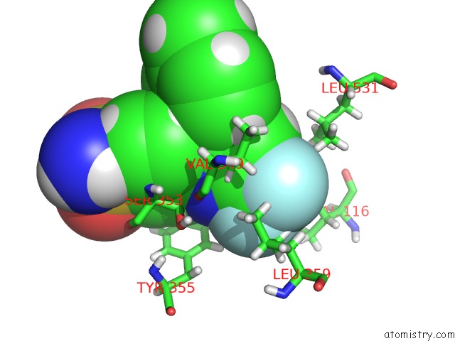
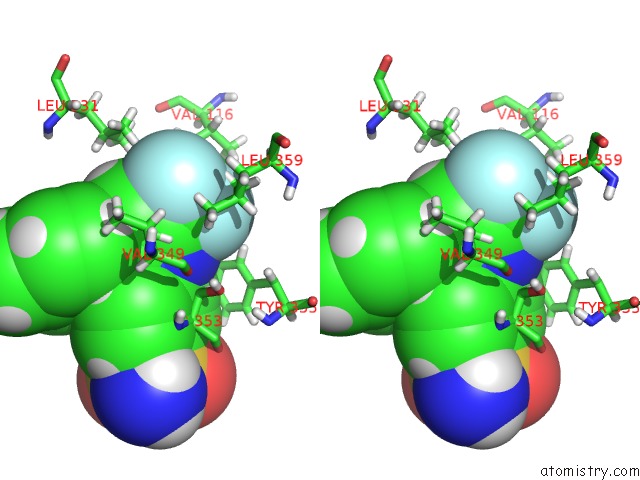
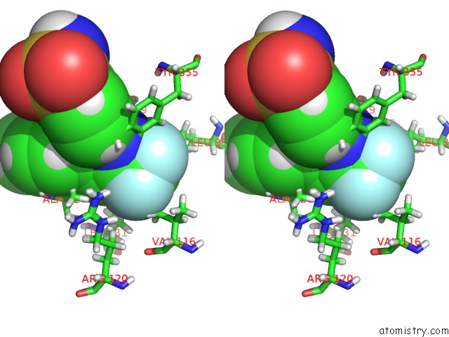
Fluorine binding site 1 out of 6 in 3kk6
Go back to
Fluorine binding site 1 out
of 6 in the Crystal Structure of Cyclooxygenase-1 in Complex with Celecoxib
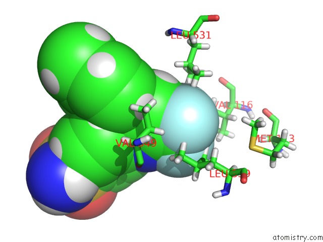
Mono view
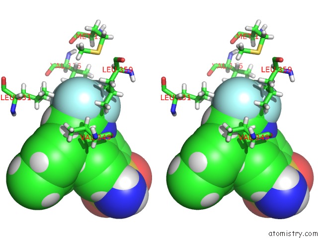
Stereo pair view
Mono view
Stereo pair view
A full contact list of Fluorine with other atoms in the F binding
site number 1 of Crystal Structure of Cyclooxygenase-1 in Complex with Celecoxib within 5.0Å range:
|
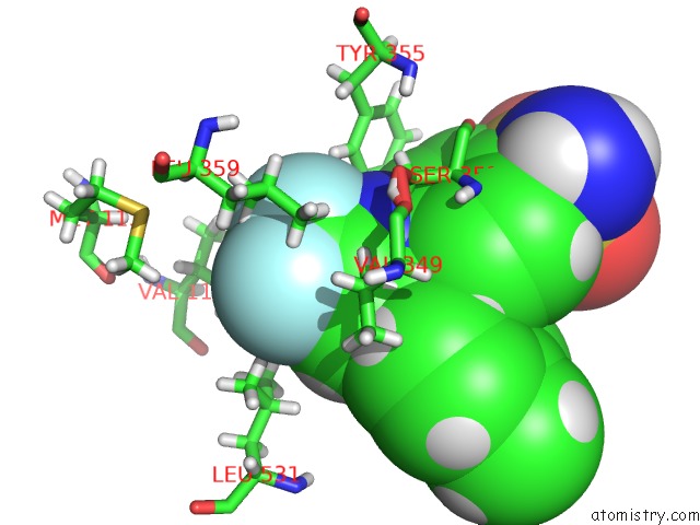
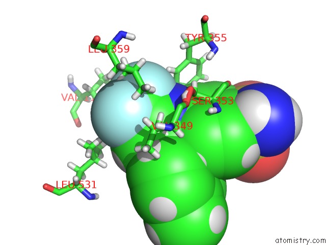
Fluorine binding site 2 out of 6 in 3kk6
Go back to
Fluorine binding site 2 out
of 6 in the Crystal Structure of Cyclooxygenase-1 in Complex with Celecoxib
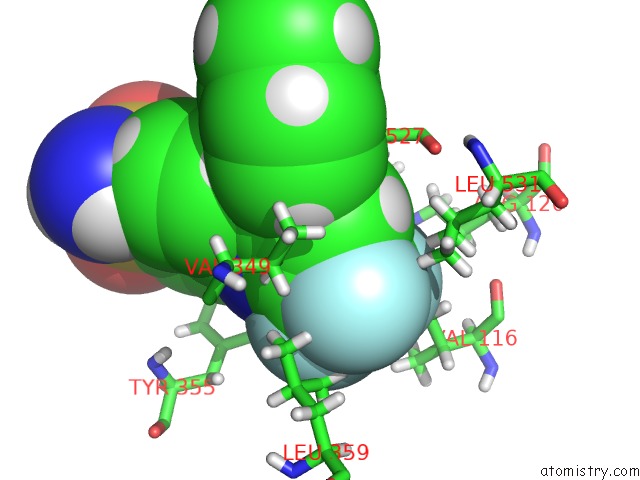
Mono view
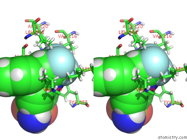
Stereo pair view
Mono view
Stereo pair view
A full contact list of Fluorine with other atoms in the F binding
site number 2 of Crystal Structure of Cyclooxygenase-1 in Complex with Celecoxib within 5.0Å range:
|
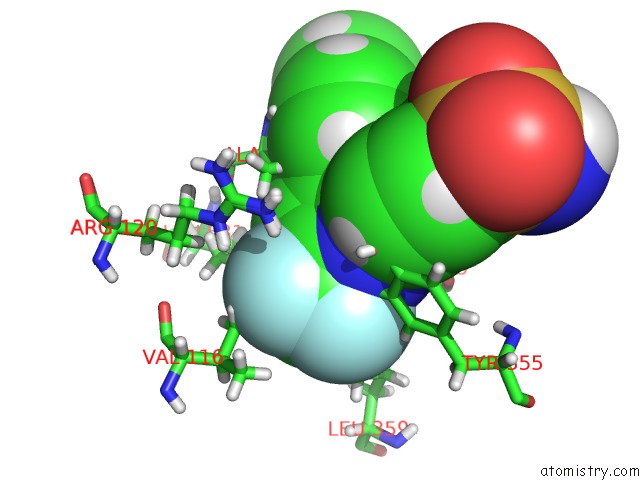
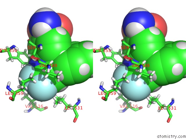
Fluorine binding site 3 out of 6 in 3kk6
Go back to
Fluorine binding site 3 out
of 6 in the Crystal Structure of Cyclooxygenase-1 in Complex with Celecoxib
Mono view
Stereo pair view
Mono view
Stereo pair view
A full contact list of Fluorine with other atoms in the F binding
site number 3 of Crystal Structure of Cyclooxygenase-1 in Complex with Celecoxib within 5.0Å range:
|
Fluorine binding site 4 out of 6 in 3kk6
Go back to
Fluorine binding site 4 out
of 6 in the Crystal Structure of Cyclooxygenase-1 in Complex with Celecoxib
Mono view

Stereo pair view
Mono view
Stereo pair view
A full contact list of Fluorine with other atoms in the F binding
site number 4 of Crystal Structure of Cyclooxygenase-1 in Complex with Celecoxib within 5.0Å range:
|
Fluorine binding site 5 out of 6 in 3kk6
Go back to
Fluorine binding site 5 out
of 6 in the Crystal Structure of Cyclooxygenase-1 in Complex with Celecoxib
Mono view
Stereo pair view
Mono view
Stereo pair view
A full contact list of Fluorine with other atoms in the F binding
site number 5 of Crystal Structure of Cyclooxygenase-1 in Complex with Celecoxib within 5.0Å range:
|
Fluorine binding site 6 out of 6 in 3kk6
Go back to
Fluorine binding site 6 out
of 6 in the Crystal Structure of Cyclooxygenase-1 in Complex with Celecoxib
Mono view
Stereo pair view
Mono view
Stereo pair view
A full contact list of Fluorine with other atoms in the F binding
site number 6 of Crystal Structure of Cyclooxygenase-1 in Complex with Celecoxib within 5.0Å range:
|
Reference:
G.Rimon,
R.S.Sidhu,
D.A.Lauver,
J.Y.Lee,
N.P.Sharma,
C.Yuan,
R.A.Frieler,
R.C.Trievel,
B.R.Lucchesi,
W.L.Smith.
Coxibs Interfere with the Action of Aspirin By Binding Tightly to One Monomer of Cyclooxygenase-1. Proc.Natl.Acad.Sci.Usa V. 107 28 2010.
ISSN: ISSN 0027-8424
PubMed: 19955429
DOI: 10.1073/PNAS.0909765106
Page generated: Wed Jul 31 19:55:20 2024
ISSN: ISSN 0027-8424
PubMed: 19955429
DOI: 10.1073/PNAS.0909765106
Last articles
Zn in 9MJ5Zn in 9HNW
Zn in 9G0L
Zn in 9FNE
Zn in 9DZN
Zn in 9E0I
Zn in 9D32
Zn in 9DAK
Zn in 8ZXC
Zn in 8ZUF