Fluorine »
PDB 3kqn-3l8s »
3kr2 »
Fluorine in PDB 3kr2: Crystal Structure of Hpnmt in Complex Adohcy and 5-Fluoro-1H- Benzo[D]Imidazol-2-Amine
Enzymatic activity of Crystal Structure of Hpnmt in Complex Adohcy and 5-Fluoro-1H- Benzo[D]Imidazol-2-Amine
All present enzymatic activity of Crystal Structure of Hpnmt in Complex Adohcy and 5-Fluoro-1H- Benzo[D]Imidazol-2-Amine:
2.1.1.28;
2.1.1.28;
Protein crystallography data
The structure of Crystal Structure of Hpnmt in Complex Adohcy and 5-Fluoro-1H- Benzo[D]Imidazol-2-Amine, PDB code: 3kr2
was solved by
N.Drinkwater,
J.L.Martin,
with X-Ray Crystallography technique. A brief refinement statistics is given in the table below:
| Resolution Low / High (Å) | 45.72 / 2.30 |
| Space group | P 43 21 2 |
| Cell size a, b, c (Å), α, β, γ (°) | 94.426, 94.426, 188.185, 90.00, 90.00, 90.00 |
| R / Rfree (%) | 21.2 / 24.7 |
Fluorine Binding Sites:
The binding sites of Fluorine atom in the Crystal Structure of Hpnmt in Complex Adohcy and 5-Fluoro-1H- Benzo[D]Imidazol-2-Amine
(pdb code 3kr2). This binding sites where shown within
5.0 Angstroms radius around Fluorine atom.
In total 2 binding sites of Fluorine where determined in the Crystal Structure of Hpnmt in Complex Adohcy and 5-Fluoro-1H- Benzo[D]Imidazol-2-Amine, PDB code: 3kr2:
Jump to Fluorine binding site number: 1; 2;
In total 2 binding sites of Fluorine where determined in the Crystal Structure of Hpnmt in Complex Adohcy and 5-Fluoro-1H- Benzo[D]Imidazol-2-Amine, PDB code: 3kr2:
Jump to Fluorine binding site number: 1; 2;
Fluorine binding site 1 out of 2 in 3kr2
Go back to
Fluorine binding site 1 out
of 2 in the Crystal Structure of Hpnmt in Complex Adohcy and 5-Fluoro-1H- Benzo[D]Imidazol-2-Amine
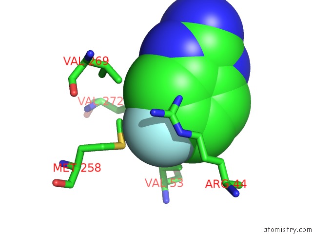
Mono view
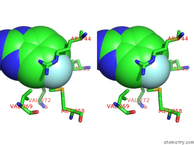
Stereo pair view
Mono view
Stereo pair view
A full contact list of Fluorine with other atoms in the F binding
site number 1 of Crystal Structure of Hpnmt in Complex Adohcy and 5-Fluoro-1H- Benzo[D]Imidazol-2-Amine within 5.0Å range:
|
Fluorine binding site 2 out of 2 in 3kr2
Go back to
Fluorine binding site 2 out
of 2 in the Crystal Structure of Hpnmt in Complex Adohcy and 5-Fluoro-1H- Benzo[D]Imidazol-2-Amine
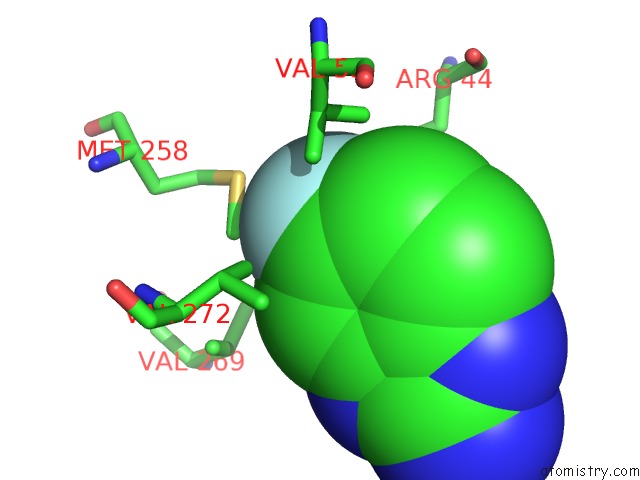
Mono view
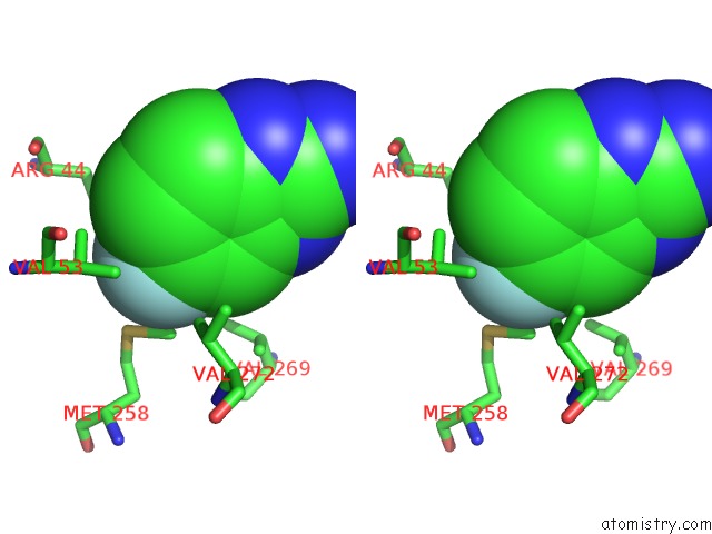
Stereo pair view
Mono view
Stereo pair view
A full contact list of Fluorine with other atoms in the F binding
site number 2 of Crystal Structure of Hpnmt in Complex Adohcy and 5-Fluoro-1H- Benzo[D]Imidazol-2-Amine within 5.0Å range:
|
Reference:
N.Drinkwater,
H.Vu,
K.M.Lovell,
K.R.Criscione,
B.M.Collins,
T.E.Prisinzano,
S.A.Poulsen,
M.J.Mcleish,
G.L.Grunewald,
J.L.Martin.
Fragment-Based Screening By X-Ray Crystallography, Ms and Isothermal Titration Calorimetry to Identify Pnmt (Phenylethanolamine N-Methyltransferase) Inhibitors. Biochem.J. V. 431 51 2010.
ISSN: ISSN 0264-6021
PubMed: 20642456
DOI: 10.1042/BJ20100651
Page generated: Wed Jul 31 20:12:40 2024
ISSN: ISSN 0264-6021
PubMed: 20642456
DOI: 10.1042/BJ20100651
Last articles
Zn in 9J0NZn in 9J0O
Zn in 9J0P
Zn in 9FJX
Zn in 9EKB
Zn in 9C0F
Zn in 9CAH
Zn in 9CH0
Zn in 9CH3
Zn in 9CH1