Fluorine »
PDB 3kqn-3l8s »
3l2w »
Fluorine in PDB 3l2w: Crystal Structure of the Prototype Foamy Virus (Pfv) Intasome in Complex with Manganese and GS9137 (Elvitegravir)
Protein crystallography data
The structure of Crystal Structure of the Prototype Foamy Virus (Pfv) Intasome in Complex with Manganese and GS9137 (Elvitegravir), PDB code: 3l2w
was solved by
S.Hare,
S.S.Gupta,
P.Cherepanov,
with X-Ray Crystallography technique. A brief refinement statistics is given in the table below:
| Resolution Low / High (Å) | 35.90 / 3.20 |
| Space group | P 41 21 2 |
| Cell size a, b, c (Å), α, β, γ (°) | 157.880, 157.880, 125.090, 90.00, 90.00, 90.00 |
| R / Rfree (%) | 19.4 / 22 |
Other elements in 3l2w:
The structure of Crystal Structure of the Prototype Foamy Virus (Pfv) Intasome in Complex with Manganese and GS9137 (Elvitegravir) also contains other interesting chemical elements:
| Manganese | (Mn) | 3 atoms |
| Zinc | (Zn) | 1 atom |
| Chlorine | (Cl) | 1 atom |
Fluorine Binding Sites:
The binding sites of Fluorine atom in the Crystal Structure of the Prototype Foamy Virus (Pfv) Intasome in Complex with Manganese and GS9137 (Elvitegravir)
(pdb code 3l2w). This binding sites where shown within
5.0 Angstroms radius around Fluorine atom.
In total only one binding site of Fluorine was determined in the Crystal Structure of the Prototype Foamy Virus (Pfv) Intasome in Complex with Manganese and GS9137 (Elvitegravir), PDB code: 3l2w:
In total only one binding site of Fluorine was determined in the Crystal Structure of the Prototype Foamy Virus (Pfv) Intasome in Complex with Manganese and GS9137 (Elvitegravir), PDB code: 3l2w:
Fluorine binding site 1 out of 1 in 3l2w
Go back to
Fluorine binding site 1 out
of 1 in the Crystal Structure of the Prototype Foamy Virus (Pfv) Intasome in Complex with Manganese and GS9137 (Elvitegravir)
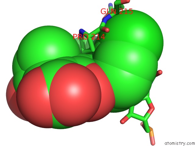
Mono view
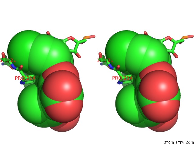
Stereo pair view
Mono view
Stereo pair view
A full contact list of Fluorine with other atoms in the F binding
site number 1 of Crystal Structure of the Prototype Foamy Virus (Pfv) Intasome in Complex with Manganese and GS9137 (Elvitegravir) within 5.0Å range:
|
Reference:
S.Hare,
S.S.Gupta,
E.Valkov,
A.Engelman,
P.Cherepanov.
Retroviral Intasome Assembly and Inhibition of Dna Strand Transfer Nature V. 464 232 2010.
ISSN: ISSN 0028-0836
PubMed: 20118915
DOI: 10.1038/NATURE08784
Page generated: Mon Jul 14 17:40:43 2025
ISSN: ISSN 0028-0836
PubMed: 20118915
DOI: 10.1038/NATURE08784
Last articles
Fe in 2YXOFe in 2YRS
Fe in 2YXC
Fe in 2YNM
Fe in 2YVJ
Fe in 2YP1
Fe in 2YU2
Fe in 2YU1
Fe in 2YQB
Fe in 2YOO